INTRODUCTION
There is an under reporting and under appreciation of the anatomical relationship of the chest to the cervical spine, head and shoulder. This case report demonstrates this relationship and a resolution of a chronic case of torticollis. A review of the literature indicates these anatomical relationships are ignored.
Torticollis is defined as “a stiff neck or wry neck, caused by spasmodic contractions of neck muscle drawing the head to one side with the chin pointing to the other side. Most often involving the sternocleidomastoid muscle unilaterally.” It can be congenital or acquired.1
This case report draws on the anatomical relationship that can be described as “Origins and Insertions”.2 The “wry muscle” are the scalenes and the sternocleidomastoid (SCM). Their involvement, resulting in torticollis, is involving spinal cord structures.3 Muscle contraction is caused by neurological input.4 The SCM’s and Scalenes 's response to the mechanoreceptors distress in the tendons of their origin and insertions is to contract. The adjusting of the ribs reduced the “noxious stimuli.”
This report introduces the Rib Cage Compression Test, which reveals the rib cage involvement in neck pain and ROM issues, and the Shoulder Squeeze Test, which discerns the SCM involvement in patient complaints. Adjusting protocols are explained and other issues discussed such as managing chronic rib/chest wall subluxations including activities of daily living, i.e.: how she puts a bra on, reaching behind her with her right arm is to be avoided. Work limitations are discussed, as are recreational activities.
CASE REPORT
The patient was born after a 12-hour stalled labor and Caesarean section. She was diagnosed with torticollis from birth trauma. She was suffering with this condition when she presented in February of 2021. She had regular contact with providers through out her life. All prescribed “routine” stretching that resulted in headaches and soreness and no change in her condition.5–8 I mention this as an indication she was receiving the standard of care. All of her previous providers had recommended stretching of the affected muscles, yielding no improvement, and neglecting the injured chest joints.
Physical Examination
The patient held her head and cervical spine in right lateral flexion. She had difficulty straightening her head and had no ability to turn her head to the left.1 She complained of pain in the right side of her neck and head of a persistent nature. She had pain on palpation of the right sternal joints, the edge of the sternum being palpable. Axillary palpation on the right was painful to the touch. All was normal on the left. She had a kyphotic cervical spine with history of 2 motor vehicle accidents (MVAs) and athletic injuries (mostly soccer) with low back and knee complaints. We performed the Rib Cage Compression Test and she demonstrated improved cervical ROM and pain relief. She also demonstrated hypermobile joint syndrome, a benign syndrome including joint laxity.9
Diagnosis
Her diagnosis was subluxation of the first through fourth costosternal joints on her right side, including the right sternoclavicular joint. The neuromuscular response to this set of unresolved subluxations caused a persistent tightening (guarding) of the scalenes group and sternocleidomastoid muscle.4 This resulted in an inability to turn her head to the left, her head to be tilted to the right and produced a constant headache at the SCM insertion on the lateral skull.2 This created torticollis.1
Treatment
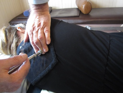
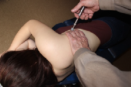
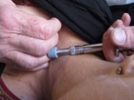
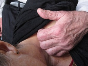
Treatment included gentle adjusting of the right proximal clavicle and first four ribs, which were posterior and lateral to the sternum.9 See Figures 1-4.
The result was an immediate dramatic increase in cervical range of motion, i.e. chin over left shoulder with ease or the first time in her life. This condition has not returned.
Management
Her continued care was focused on the other issues she was experiencing. Later, the intervention included gentle adjusting of the cervical spine (kyphotic curve) and introduction of a 2" rib belt to help stabilize the upper chest region. (See below). This was to manage her other complaints, the torticollis was resolved. Within 3 weeks she was asymptomatic and her treatment plan was focusing on management of the other injuries. Since she had been living with this condition since birth, she had never thought about why she had pain or what she was doing to exacerbate it. Only with pain relief and reoccurring onset of pain can one start to recognize which patterns of behavior have an adverse effect on the resolution of the injury. In her case, lifting and carrying things for work was a major contributing factor, but day to day activities like housekeeping, shopping and grooming also had to be approached differently, more ergonomically mindful of the right chest injury. We performed the rib cage compression test to discern the role the ribs’ subluxations have on affecting the cervical pain and ROM. She had significant relief from the adjusting, but additional compression of the ribs gave her further relief.10
Treatment For Subluxated Ribs
Rib compression adjusting can be done with an adjusting instrument (such as a spring loaded adjusting instrument). If the doctor is much smaller than the patient, the patient can be seated and, if needed, only 1 side at a time is treated. If the patient is in a hospital bed or wheelchair, perform the adjustment 1 side at a time.


When correcting the posterior rib subluxation, cross the arm over the patient’s chest to move the scapula away from the angle of the ribs. (See Figure 1)
Place the adjusting instrument over the doctor’s contact fingers and tap repeatedly. Note: This adjustment can be used through out the ribs. (See Figures 1 and 2)
During the initial office visit, after assessing the clavicle and rib cage involvement in this patient’s complaints, the proximal clavicle joint and ribs were adjusted. She immediately demonstrated full range of motion to the left, chin over left shoulder. Suggesting that the guarding patterns had been relieved when the rib joints were realigned. It was now necessary to determine if the ribs would stay in place with normal weight-bearing activity, such as walking. If the guarding pattern and the rib cage subluxation pattern start to return after a few minutes of walking, then driving and other activities could also cause the ribs to go back out of place, sometimes with just the weight of the arm and shoulder. On palpation of the ribs we found that she lost some of her alignment with walking. This was determined by palpation and return of her flinch response.


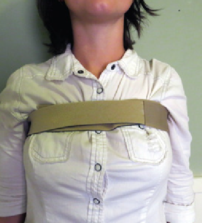
At this point, the patient was fitted with a simple rib brace (Figure 5) and asked to walk around for a few minutes. When asked whether she felt better with or without the rib belt, she stated that she felt relief with it on and wanted to wear the brace. She had no flinch response and rib alignment was maintained. This finding is consistent with the fact her arms and shoulders weight about 20 pounds apiece. Just their weight and gravity would be provocative to the injured joints. She is using the brace prophylactically for housekeeping, gardening and laundry activates.10
Managing The Rib Cage Injury
Managing a rib cage injury that is restricting arm function or creating entrapment syndromes needs to be fully understood by the patient. The more the patient can see these relationships, the more the patient can assist in managing their own injuries. Using the rib belt in this case has been essential to resolving the case. It was apparent in post-adjusting evaluation the motion and weight of her arm pulled the ribs back out of place. To allow the injured ligaments to heal they need to be supported in the position of relief.10 So the use of the rib belt was immediately apparent to her when it was removed. There was a tightening of her neck and shoulder muscles without the belt. How she dresses, including bras, sleeping positions and car behavior, (no reaching into the back seat with right arm, no pushups, bench presses or downward dog in yoga) all needed to be addressed as well as ceasing all stretching she was instructed to do previously.
Outcome
The patient had immediate relief of her chief complaint after the first intervention. There was no return of limited cervical ROM and no head tilt. The issue was the management of a chronic rib cage injury. At the time of presentation she worked for a small production company requiring her to transport and move material. Her next job was as a special education teacher with three different classrooms on 3 different campuses. There was a lot of moving educational supplies. Now she is in graduate school with a backpack and a grueling study schedule. She is no longer plagued by headaches or restricted ROM. She has regular care to her cervical spine, ribs, low back and knee complaints (avoiding soccer helps). She still has to be mindful of her ergonomics. The rib belt should be used when stressing the shoulder and chest. Heavy lifting, gardening and housekeeping have caused set backs for neck pain and stiffness, but no return of the head tilt or restriction of cervical motion.
DISCUSSION
This patient and her mother can testify that they spared no efforts to resolve her birth-related injuries. Out of more than 30 providers over the 20 + years of searching, no one mentioned the words “ribs” or “chest” to either of them. The scalenes and SCM muscles attach to the chest. Disruption of the chest joints can cause a reflex guarding to protect the remaining integrity of a failing vital cavity, the chest. However current medical literature and treatment protocols fail to acknowledge this. Consequentially, they fall short of actually addressing what is causing the condition. Use of the Rib Cage Compression Test is the first step to determining the causative factors and providing long term relief to the patient. Gentle adjusting of the ribs in the direction of correction as opposed to standard thoracic manipulation is recommended.


This test has 2 purposes: The doctor needs to see how much the restriction or pain with cervical activity is related to the subluxated rib cage. The patient can see that any positive changes from having their ribs compressed reveals that the rib subluxations are part of their injury and diagnosis
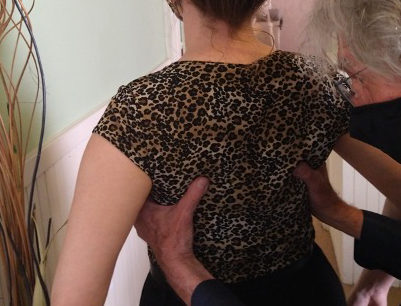
The Rib Cage Compression Test
The Rib Cage Compression Test is performed to demonstrate the effects of the injured, subluxated rib cage on cervical function, entrapment syndromes and other associated complaints.
The patient first demonstrates cervical range of motion, and then The Rib Cage Compression Test is performed. While holding the compression, the patient is asked to repeat range of motion. Any differences are noted.
In the majority of cases, this can be done with the doctor standing behind the patient. Both hands are used; one on each side at the axilla of the patient, and a squeezing or compression of the ribs from lateral to medial is performed. See Figure 6. The compression should be maintained for 30 seconds. The patient then repeats the range of motion and any changes are noted.

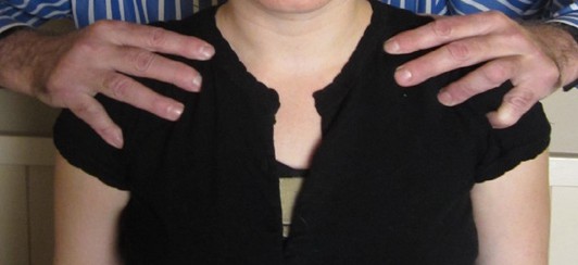
The Shoulder Squeeze Test
This test is meant to relieve the sternoclavicular joints of distress and relax the SCMs on each side. The positive finding for this test is relief of tension and possibly pain on the side of the head and neck. The SCM has in it the external jugular vein and relieving the compression of this vein can give relief to pain in the sinuses, frontal area, eyes, muffled hearing, dizziness and difficulty swallowing. This squeezing is maintained to a count of 30. (Figure 7). Both of these tests are positive if there is relief. This test will also reieve distress of the infrahyoid mucsles if difficulty in swallowing is a complaint.
CONCLUSION
Our patient had the state of the art applied to her condition.5–8 I find no reference of rib cage involvement in any article or chapter of a textbook on the subject. She was faithful, for years, dutifully stretching her tight neck (wry) muscles to the point of pain, on a regular routine. There was no improvement and continued pain, in part from the stretching, for more the 2 decades. Here is evidence that Injuries to the rib cage and sternoclavicular joints may be responsible for the scalene muscles and SCM to be tight and rigid, resulting in Torticollis.