

Introduction
Muscular dystrophy (MD) represents a group of inherited neuromuscular disorders characterized by progressive muscle degeneration, weakness, and declining functional capacity. These conditions arise from approximately 30 different forms with genetic mutations that disrupt proteins essential for maintaining muscle fiber integrity, most notably dystrophin in Duchenne and Becker muscular dystrophies.1 The absence or dysfunction of these proteins leads to sarcolemmal instability, repeated cycles of muscle damage, and eventual replacement of muscle tissue with adipose and fibrotic tissue. Despite advances in genetic and pharmacologic therapies, there remains no definitive cure for muscular dystrophy. As a result, management strategies have shifted toward preserving function, maintaining independence, and improving quality of life. Emerging evidence supports the role of appropriately dosed exercise, strength and conditioning principles, and integrative care models in achieving these outcomes.2
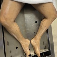
Muscular dystrophies arise from mutations affecting proteins responsible for muscle fiber stability, force transmission, and cellular repair. In many forms of MD such as Duchenne and Becker muscular dystrophy (See Figure 1, a photo of lower extremities of cadaver with MD), absent or defective dystrophin leads to sarcolemmal instability, increased susceptibility to mechanical stress, and repeated cycles of muscle damage and incomplete regeneration.3

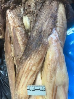
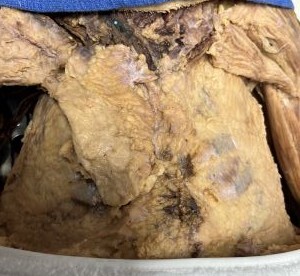
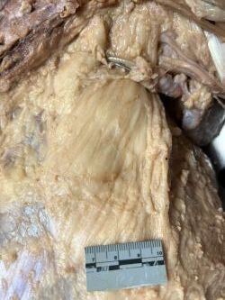
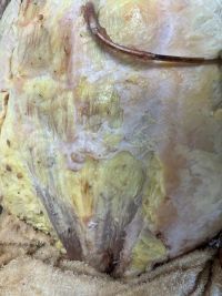
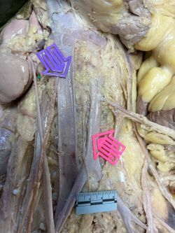
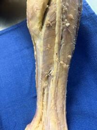
Over time the abnormalities of muscular dystrophy lead to progressive muscle fiber necrosis (as noted in the thigh muscles of cadaver with MD, see Figure 2), replacement of contractile tissue with fat and connective tissue (seen in pectoralis muscles in cadaver with MD, Figures 3 and 4, psoas major and minor muscles in cadaver with MD, Figure 5, rectus abdominis and anterior abdominal wall muscles, Figure 6, and leg and foot muscles, Figure 7), reduced force production and endurance, and altered neuromuscular control.4

.jpeg)
.jpeg)
.jpeg)
The progression of muscular dystrophy is marked not only by muscle weakness but also by secondary complications including joint contractures, postural abnormalities, altered gait mechanics, and pain. These secondary impairments often contribute significantly to functional limitations. As muscle fibers degenerate, the body compensates through altered movement patterns, placing increased stress on joints, connective tissues, and the neuromuscular system.5 Importantly, while the primary disease process is irreversible (like the fatty change and atrophy of the muscles as noted in this cadaver), secondary biomechanical and functional impairments remain modifiable, making them key targets for conservative and rehabilitative interventions. This distinction provides the foundation for integrating strength and conditioning strategies and manual therapies into patient care. Such impairments, as would be expected in this cadaveric case, are what should be homed in on to rectify in patients with a neuromuscular disease like MD.
Discussion
In individuals with muscular dystrophy, the objective in care shifts toward preserving neuromuscular function, improving movement efficiency, and maintaining independence rather than maximizing strength output. Research indicates that low-to-moderate intensity resistance training is both safe and beneficial when appropriately prescribed. A systematic review examining exercise interventions in muscular dystrophy populations found that strength training improved balance, gait, and functional capacity without significant adverse effects.6 Similarly, clinical evidence demonstrates that submaximal resistance programs can be safely implemented in individuals with Duchenne muscular dystrophy, with improvements observed in functional performance and tolerance to activity.7
Neuromuscular rehabilitation literature suggests that exercise should prioritize endurance, coordination, and movement quality over maximal strength gains.8 Notably, evidence suggests that while increases in maximal strength may be limited, functional improvements such as walking capacity, endurance, and balance are consistently observed.6
Exercise interventions in MD populations have been shown to improve gait efficiency, enhance postural stability, maintain joint mobility, and delay functional decline. Additionally, low-load, high-repetition activity may promote neuromuscular adaptations while minimizing muscle damage, providing a safe and effective approach for long-term management.9
Chiropractic care offers a unique contribution to the management of muscular dystrophy by targeting secondary musculoskeletal and biomechanical dysfunctions. Patients with MD frequently present with reduced joint mobility, spinal misalignment, compensatory movement patterns, and pain associated with altered biomechanics.10 A case report included in the literature review demonstrated that conservative chiropractic interventions, including low-force spinal adjustments, soft tissue techniques, and therapeutic exercise, resulted in improvements in range of motion, pain reduction, and functional capacity.1 While chiropractic care does not alter the underlying genetic pathology, it may enhance patient outcomes by improving joint mechanics, reducing pain, and optimizing neuromuscular input.
The strongest clinical implication emerging from current literature in the care of patients with MD is the value of a multidisciplinary, integrative approach. Exercise-based interventions and chiropractic care should not be viewed as isolated treatments, but rather as complementary components of a comprehensive care model. Commonly reported complementary therapies for MD include massage therapy, chiropractic treatments, osteopathy, and naturopathy.11 Massage therapy has been suggested to decrease inflammation and promote mitochondrial biogenesis in skeletal muscle that has been acutely damaged through exercise.12 Current literature supports the necessity of multidisciplinary care models in muscular dystrophy management, incorporating rehabilitation, exercise, and supportive therapies to optimize patient outcomes.6 Within this framework, chiropractic care can serve as a valuable adjunct by addressing mechanical and neuromuscular contributors to dysfunction.
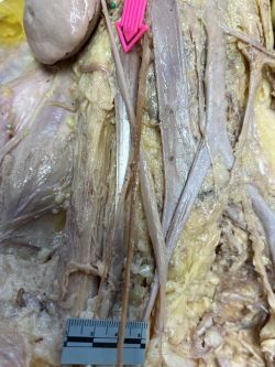
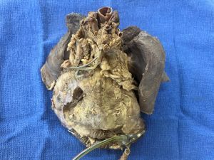
Muscular dystrophy presents complex, lifelong challenges that extend beyond skeletal muscle degeneration alone, like involvement of cardiac muscle (See Figure 8, cadaver in the anatomy lab, requiring LVAD placement due to the disorder affecting the heart) and per Kate Bushby involvement of the heart in MD can lead to cardiomyopathy and ultimately cause death13 or involvement of the pancreas with development of diabetes mellitus, as was noted in this cadaver. Also, as seen in this cadaver, the muscle in blood vessel walls can be affected resulting in decreased diameter of major blood vessels (See Figure 9, decreased caliber of the diameter of inferior vena cava and descending abdominal aorta). An additional finding in this cadaver was the presence of a micropenis, which can be a finding in certain types of congenital muscular dystrophy.
_and_descending_abdominal_aorta_(pink_arrow).jpeg)
Limitations and Future Directions
Although evidence supporting exercise interventions in muscular dystrophy continues to grow, limitations remain. Much of the literature consists of small clinical trials or systematic reviews with heterogeneous populations. Additionally, research specifically examining chiropractic care in MD is limited primarily to case reports. Future research should focus on longitudinal studies evaluating integrative care models, standardized exercise dosing protocols, objective functional outcome measures, and interdisciplinary collaboration.
Conclusion
While the primary disease process cannot currently be reversed, secondary impairments related to movement, biomechanics, and function are modifiable. Movement and biomechanics would be abnormal in muscles with marked fatty replacement and atrophy, as seen in the pectoralis, psoas, gastrocnemius, thigh, and abdominal muscles in the cadaver presented here with MD. Per Eric Knapp’s story, Spinal Column Stressology treatments by Dr. Joseph Bahan to Eric, who had MD, enabled Eric to lift a cup to his mouth, to be more flexible, and to have restoration of voluntary movements to his legs.14 A belief of Dr. Bahan is that chiropractic adjustments reduce some of the symptoms of MD.15 Emerging evidence supports the safe implementation of submaximal strength and conditioning interventions, with demonstrated improvements in functional capacity, endurance, and quality of life. When combined with chiropractic care targeting joint mechanics and neuromuscular function, these approaches offer a promising, integrative strategy for managing individuals with muscular dystrophy.16 The interdisciplinary model aligns with the evolving emphasis on function-driven care, highlighting the importance of preserving movement, independence, and overall patient well-being. The patient with MD presented in this article lived until he was 29 years old, but his quality of life might have been improved with a multidisciplinary approach to treatment, including some type of chiropractic modality.
More research is needed to study the effects of CAM (complementary alternative modalities) on neuromuscular conditions in pediatric patients, especially those with MD. CAM treatments have been reported to help decrease pain in adults with neuromuscular diseases, such as painful peripheral neuropathy.17 Also, as noted in the conclusion of the article by Kenya and Vuylya in JCC, muscular dystrophy may respond favorably to chiropractic care in conjunction with physical therapy and therapeutic exercises.1 They described significant improvement in the condition of a 23-year-old man with MD after chiropractic adjustments to different areas of the spine, physical therapy, and exercises after 12 weeks. In summary, people with muscular dystrophy deserve the very best treatment modalities to help them live a life free of as much pain as possible for as long as they can.