
INTRODUCTION
Diffuse idiopathic skeletal hyperostosis (DISH, Forestier’s Disease) is a generalized spinal and extraspinal articular disorder which is characterized by ligamentous calcification and ossification. The most common radiographic expression of this condition is anterior flowing hyperostosis involving the anterior longitudinal ligament of the cervical, thoracic and lumbar vertebra; the lower thoracic spine is the most common site of spine involvement. DISH typically involves at least 4 contiguous vertebral bodies, lacks signs of disc degeneration and spares the apophyseal joints. Patients may be asymptomatic or have complaints similar to those of degenerative joint disease: morning stiffness and low-grade musculoskeletal pain, especially of the spine and related articulations.1 This condition is more frequent and more severe in males throughout life, and the incidence of the disease increases with age. There is a higher prevalence in Caucasian populations than in Korean or black Africans.2 The precise cause is undetermined; however recent studies suggest it is associated with a greater body mass index (obesity), higher serum uric acid levels, type 2 diabetes mellitus, impaired lipid metabolism, hypertension, metabolic syndrome and an increased risk for cardiovascular diseases. Serum matrix Gla protein (a vitamin K dependent protein) may be a marker of osteometabolic syndromes that cause hyperostosis. Genetic factors human leukocyte antigen (HLA) and collagen type VI alpha 1 (COL6A1) genes, environmental exposures (fluoride, vitamin A/retinol), and drugs (isotretinoin, etretinate, acitretin, other vitamin A derivatives and teriparatide) may be associated with DISH.2–6 Fractures from low-energy trauma with neurological deficit are more common in patients with DISH than in the general population and the incidence of fractures with complications and increased mortality are associated with the elderly. Delayed diagnosis of spinal fractures is common when neurological deficits are absent.2,7–9 Conventional treatment is directed toward symptoms of pain and stiffness with analgesics and anti-inflammatories. Surgical intervention is used to resolve spinal fractures presenting with neurological deficit.7–9 Limited literature documentation of DISH etiology and spine manipulation of its symptoms exists.
CASE REPORT
We report the treatment of a patient who complained of very sharp pain from the lower spine to the left hip and from the left hip to the pubic bone and numbness of the left anterior thigh. Application of the Cox Technic lumbar spine protocol was used to treat his hip and groin pain. The pathogenesis of DISH, the case history of the patient, examination findings, diagnostic imaging, treatment and conclusions are described.
The patient was a 69-year-old male, who stood 5’9" tall and weighed 162 lbs. His gait was guarded as he avoided putting full weight on his left hip, and he exhibited a slight forward antalgic lean. Lumbar ranges of motion were limited in flexion 45/90, extension 10/30, left lateral flexion 5/20, right lateral flexion 15/20 and rotation bilaterally 20/30. Hip range of motion was normal, with low back pain produced at the end range of flexion and lateral rotation. Straight leg raise, double leg raise and Valsalva’s maneuver were negative for disc displacement producing radicular pain. Kemp’s test produced low back and iliac crest pain on the left. Patrick FABERE test was negative. Lower extremity reflexes were normal; however, hypoesthesia was noted on his left anterior/lateral thigh (upper lumbar dermatomes). Palpation showed spasm and hypersensitivity of the erector spinae, quadratus lumborum muscles bilaterally and gluteal muscles and tensor fascia lata on the left.
Diagnostic Imaging
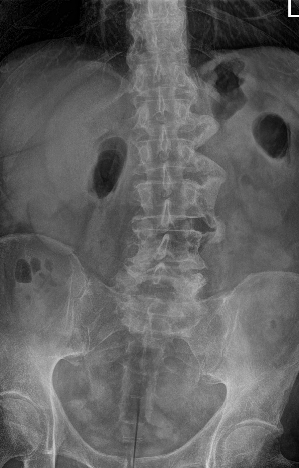
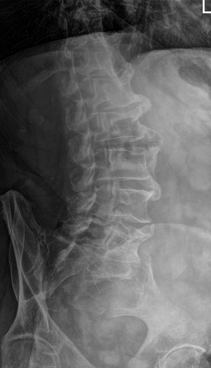
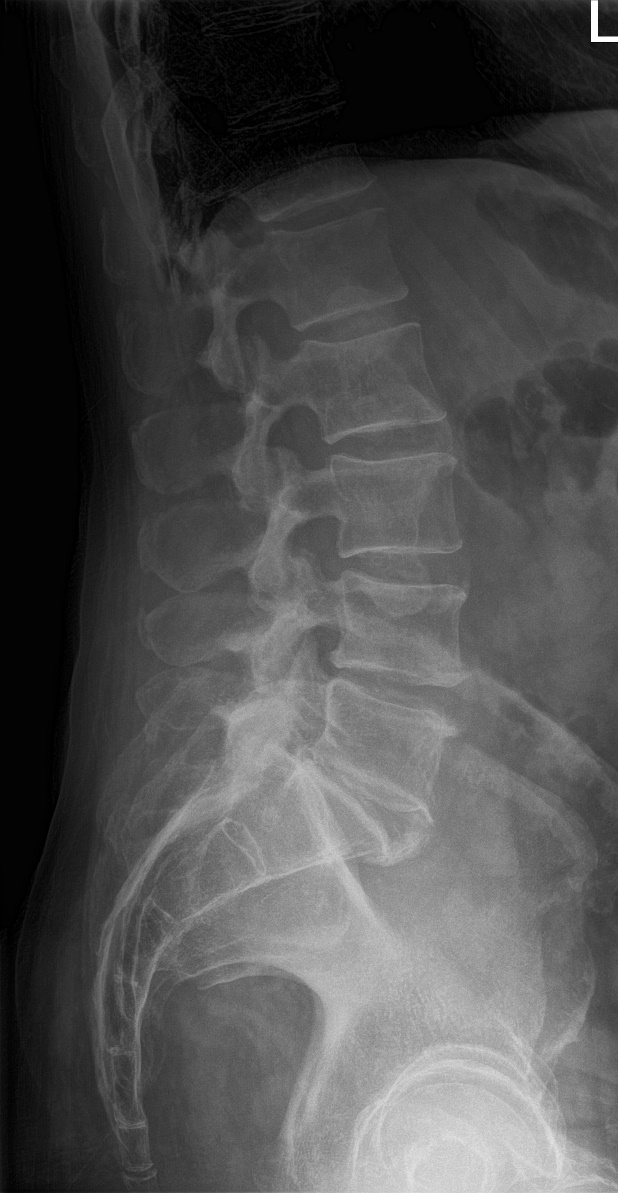
Plain film x-rays of the lumbosacral spine with obliques demonstrated flowing left-sided ligament ossification with ankylosis extending from L1-L4 with preservation of disc height, mild-to-moderate left facet arthrosis at L5-S1 and anterior ligament ossification ankylosis of the L5-S1 disc space, mild levoscoliosis, apex at L3-L4 and low left hemipelvis. See Figures 1 to 4.


Treatment
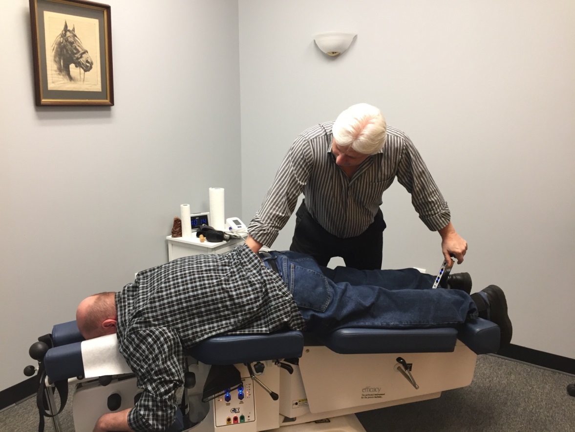
The treatment plan was to use Cox Technic Protocol II from T12 to L4. See Figure 5. Protocol II (long y axis distraction with segmental spinal mobilization) is chosen because the patient did not exhibit any radicular symptomatology. Prior to treatment, tolerance testing was performed as well. Tolerance testing involves gentle long-y axis distraction of each spinal segment from L5 to T12 before starting flexion distraction spinal manipulation Protocol II. With Protocol II, all ranges of motion are performed as necessary to regain optimum motion to the spinal joints (flexion, lateral flexion, circumduction, extension). On tolerance testing, left lateral flexion aggravated the patient’s lower back, hip and thigh pain, so treatment consisted of 3 applications of flexion, right lateral flexion and y-axis distraction with manual massage of the Bladder Channel points along the lumbar spine and the muscles of the hip and buttock. The patient stated that y-axis distraction seemed to be the most effective in reducing the tightness and pain in his lower back, hip and thigh. Treatments were administered 2 times per week. The patient was encouraged to continue the stretches and core exercises he had started prior to seeking treatment. Chondroitin sulfate and hyaluronic acid were prescribed twice daily at 2000 mg and 200 mg, respectively.

DISCUSSION
The effectiveness of spinal manipulation for the relief of segmental dysfunction and pain associated with DISH requires further study and documentation. Nutritional benefits also need further documentation. Treatment did include 2000 mg of chondroitin sulfate and 200 mg of hyaluronic acid twice per day to reduce inflammation and pain and perhaps slow degenerative changes.10–19 Painful human intervertebral discs exhibit nerve growth deep into the intervertebral disc (IVD) from the dorsal root ganglion. Chondroitin sulfate (glycosaminoglycan) inhibits growth of the dorsal root ganglion axons into the IVD. Intact glycosaminoglycans such as chondroitin sulfate secreted from notochordal cells are potential candidates that could be useful to reduce neurite growth in painful IVDs.20
Cox Flexion-Distraction is an effective treatment for lumbar and thoracic spine conditions.21 There is a published protocol algorithm for its application.22 Flexion distraction is 1 of the 3 most studied adjustive procedures.23 Its forces are able to be measured in real-time and to be taught.24,25 It is an effective method for the treatment of spinal pain disorders arising from disc herniations [contained22,26–29 and non-contained21,23,28,30] and other spinal motor unit dysfunctions like lumbar spinal stenosis,31–36 spondylolisthesis22,30,37,38 [even a dual level38], synovial cyst,39–42 sciatica,28,43–45 pregnancy-related sciatica,46 spine-related testicular pain,47 adjacent segment disease and post-surgical continued pain,48–51 etc.22,28 Compared to physical therapy active exercise, flexion distraction showed significantly better outcomes for patients with lumbar radiculopathy.52–54 A series of pre and post MRI studies of cervical and lumbar spine disc herniations showed reduction in size of the herniation after flexion distraction treatment.55,56 A study of the spinal reflex excitability changes after flexion distraction showed how trunk flexion is joined by inhibition of the motor neuron pool.57 The erector spinae muscles’ resting rate and contraction ability are positively affected with flexion distraction therapy.58 Flexion distraction affects muscles and nerves affected by spinal pain conditions, disc and non-disc related.
In Traditional Chinese Medicine, the Bladder Channel begins at the inner canthus of the eyes, travels across the top of the head and descends paraspinally to the buttocks and continues down the posterior thigh and leg (following the path of the sciatic nerve), passes behind and under the lateral malleolus and terminates at the lateral corner of the nail bed of the 5th toe. Trauma to the lower back will produce an obstruction of the flow of Qi and Blood in the area resulting in pain, stiffness and muscle spasm referred to as Painful Obstruction Syndrome.59 Manipulation of the local Bladder Channel points in the low back help reestablish the flow of Qi and Blood. As part of the Cox protocol this technique provides another method of resolving a painful low back condition through non-invasive, conservative means.
While DISH did not involve disc herniation in this case, the spinal motor units are affected by the calcification and ossification of the anterior longitudinal ligament. The distraction and mobilization of each vertebral segment by the application of Cox Technic allows for the optimization of spinal motor unit function and alleviates the mechanical stress which potentiates pain. A case of DISH with chronic neck pain treated with Cox manual cervical distraction resulted in a decrease in pain severity and frequency and improved her ability to perform activities of daily living.60 This case shows flexion-distraction to have helped treatment for hip, groin pain and thigh pain found in conjunction with DISH in this patient.
LIMITATIONS
A search of PubMed and Medline databases found few studies about the non-surgical treatment of DISH. Four studies delineating chiropractic treatment of DISH from 1995-2012 were found, 1 study using exercise therapy for lumbar DISH and 2 studies of manual treatment of cervical DISH. Further case studies are needed to help validate spinal manipulation benefits in the treatment of scleratogenous and dermatogenous low back, pelvic, thigh and lower extremity pain. We cannot state that the spinal manipulation delivered to this patient was the single reason for pain relief. Exercise and nutrition along with ergonomic treatment was included in the treatment. Questions such as: What is the neurophysiological explanation for the relief of this patient’s pain following spinal manipulation? How do we know more or less visits would have resulted in improved clinical relief? Would the patient have recovered with no treatment? The purpose of this case study is to bring awareness and further study of the clinical cause of pain prescribed in this case with DISH.
CONCLUSION
This case describes flexion distraction spinal manipulation of the lumbar and lower thoracic spine in a patient diagnosed with DISH for the relief of left hip, groin and anterior thigh pain. Four treatments decreased the numeric pain scale rating from 10 to no thigh or hip pain and at 14 treatments to no pain in the low back, hip, groin or anterior thigh. Exacerbation of left anterior thigh pain following gardening prompted repeated spine manipulation.
As the incidence of obesity and Diabetes mellitus increases, so will the incidence of DISH. Chiropractors should be weary of the potentially serious complications of this condition when seemingly minor trauma is part of an elderly patient’s clinical history. Flexion distraction spinal manipulation may offer a viable treatment option for patients with DISH.
ACKNOWLEDGEMENTS
Thank you to Dr. James Cox and Julie Cox-Cid for help in manuscript preparation.