Introduction
Nephrolithiasis is the development of kidney calculi or kidney stones (KS) in the kidney. A KS is a hard crystalline mineral formed in the kidneys.1 According to The National Health and Nutrition Examination Survey,2 prevalence of kidney stone disease (KSD) has seen an upward trend in the general population over 30 years, with the rise from 1 in 11 from 1 in 20 persons since the 1990’s. The highest KS prevalence is in males ages 49–59, at 12.6%, with 17.8% in males older than 60. KS occurs in females ages 20-39 at a rate of 7.5% and with the trend rising in females over 60 to 9.8%.3–5 The prevalence of recurrence in the global population for those with a history of KSD is between 32 -50%.1,4,6,7 The economic impact of KSD on the United States population can exceed $5 billion (about $15 per person in the US) including emergency department visits, hospital admissions and lost work productivity.8
KS forms in the kidney in varying chemical compositions and sizes and can become lodged in the renal calyces or renal pelvis (nephrolithiasis). The classic pain presentation often includes pain in the low back and sacroiliac joint with radiating pain into the posterior-lateral thigh, pelvis, and groin area.1,9,10 Clinical presentations of symptoms are often similar to mechanical low back pain (LBP) and sacroiliac joint pain (SJP). Low back pain and SJP are common symptoms that often lead a patient to consult a chiropractic healthcare provider.11
Although it is uncommon for patients with acute visceral pathologies to seek out the chiropractor prior to seeing their family practitioner, it is of utmost importance that chiropractors are aware that visceral clinical symptoms can be similar to mechanical injury pathologies.12,13
The following case report discusses the diagnosis of atypical nephrolithiasis. The patient had left-sided pain in the mid-thoracic spine and ribs, lower back pain, and SJP. Although the patient reported LBP and SJP no posterior-lateral thigh, pelvis or groin pain was noted. Mid-thoracic pain is also atypical for nephrolithiasis. Few chiropractic articles14 have reported nephrolithiasis presenting as mechanical LBP and SJP with temporary relief of symptoms responding to chiropractic manipulation. There is a paucity of literature on identifying spinal signs of pain between non-musculoskeletal or visceral pain and musculoskeletal sources15
Case Report
Clinical History
A 63 -year-old female patient had been seen in the Palmer College teaching clinic in previous years for LBP, SJP and thoracic posterior rib pain. She successfully received chiropractic manipulative therapy and mechanical massage. Her history included 1 natural childbirth and 2 Caesarean sections. She had a previous kidney stone 5 years earlier that passed after 24 hours where she experienced the typical urolithiases pain radiating into her posterior flank, pelvis, and groin. She visited the emergency department (ED) during the episode and was pain medication. She then was released from the ED to wait for the stone to pass. The patient reported rare alcohol consumption and no tobacco use and no prescribed medications.
She sought chiropractic care for LBP, SJP and mid-thoracic pain. No injury or trauma was reported but an increase in physical activity was reported by her. The presentation suggested mechanical back pain and an examination was performed. During the examination, her low back and sacral joint pain were dull in quality and constant. The mid-thoracic pain was increasing and becoming more colicky in nature. After the examination, she was sent for x-rays.
Examination
Respiration, blood pressure, and temperature were within normal limits.
There was decreased active motion in the left lumber and sacroiliac regions with pain in the end range of extension. There was decreased extension and right rotation of the lumbar spine on a sitting Kemps test. S-I hyperextension (Gaenslen’s) test was performed with left-side testing positive, a side iliac compression test, and a sacral-iliac distraction test elicited left sacral iliac pain. A straight-leg raise, and slump test were performed with negative findings on both tests. Hypertonicity was noted in the left quadratus lumborum muscle and range of motion was decreased on extension.
The palpation of left thoracic 6, 10, and 12 and associated ribs elicited pain.
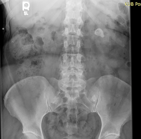
After the examination was performed, the patient was sent to the chiropractic clinic radiology department for a series of spinal views and a KUB radiograph. The KUB view (figure 1) showed a 2.6 cm (about 1.02 in) calcification within the left renal calculus.
Outcomes
The patient was referred to a urologist for an abdominal computerized tomography (CT) scan. revealing a 2.6 cm (about 1.02 in) stone in the upper pole of her left renal calyces. A urinalysis confirmed blood in the urine with high specific gravity. She was scheduled for surgery due to the size of the stone. Lithotripsy is an effective treatment suited for KSD under 2 cm, whereas stones that are larger than 2 cm (about 0.79 in) are candidates for surgery.16 The surgery removal determined the stone type was calcium oxalate. Following surgery, the patient reported no LBP, SJP or mid-thoracic pain.
_left_renal_calculus.png)
Discussion
According to The National Health and Nutrition Examination Survey2 KSD is on the rise impacting not only the physical well-being of 10.1% of US patients but adding to the economic burden at an annual cost estimated US $ 2.81billion. The rise of KSD in the population has been attributed to the prevalence of increase in diabetes mellitus, obesity, hypertension, chronic low back pain, lifestyle choices, and chronic conditions.1,17 According to researchers’3,4 understanding, the causative comorbid conditions and gender differences in KSD, (with men being twice as likely to have KSD compared to women) is crucial. It is important to note that older women are closing the gender KSD gap. These findings can alert the healthcare provider to ask questions about past KSD.
Once the stone has passed or has been surgically removed, prevention can be the key to stopping a recurrence of KSD. As Mirmozaffari18 stated, an expert computer system can be used to assist a clinician by efficiently asking questions that may show patterns of risk of KSD. After a history and physical examination of the patient’s symptoms, nephrolithiasis diagnosis is usually suspected. Researchers noted that plain film kidney, ureter, bladder (KUB) and ultrasound (USS) should be used on suspected cases of stone disease.19,20 Confirmation of the diagnosis can be made with KUB radiographs, although CT is recognized as the choice for diagnostic imaging. Kluner21 is quoted as stating, “A major disadvantage of CT, however, is the higher radiation exposure of the patient compared with KUB or IVU especially the gonads, which receive a higher radiation dose”.
Due to the prevalence and recurrence of KSD in the population, healthcare providers can direct patients in conservative self-care instructions primary to and secondarily after a kidney stone episode.2,22,23 Obesity, hyperlipidemia, diabetes, and hypertension are clinical risk factors for KSD,6,24 and these metabolic factors should be monitored. Preventive options include dietary recommendations such as increased hydration, decreased ingestion of sodium and animal proteins and an increase intake of vegetables and fruit rich in potassium.9,25,26
Nephrolithiasis is usually identified clinically due to its classic symptoms. Most patients with nephrolithiasis have renal colic and the pain can be located to the area where the stone is located.12,14,27
This case was unique in that the patient exhibited a clinical presentation of mechanical joint pain and the short-term resolution of the pain through chiropractic manipulation. LBP and SJP can share similar presenting symptoms of mechanical sources and nephrolithiasis symptoms, but the addition of mid-thoracic pain can be an atypical presentation of nephrolithiasis. The initial orthopedic evaluation reproduced mechanical sources and was used for 3 weeks. Short-term resolution each week for the LBP and SJP required use of chiropractic manipulation as a therapy.28 Resolution of the subsequent thoracic symptoms was achieved by surgical intervention to remove the KS.
Limitations
This study is a case report with data collected from medical records and supplemented by patient recall of the event. This is a single-patient case with atypical presentation of nephrolithiasis, with the unique short-term positive response to spinal manipulation. These are not the typical symptoms of nephrolithiasis compared to other cases.
Conclusion
This case shows that nephrolithiasis can imitate mechanical lesions such as LBP and SJP. Pain in the mid-thoracic area is uncommon in nephrolithiasis. Clinicians need to be cognizant of patients presenting with LBP, SJP, flank and thoracic pain for not only mechanical injury but also for signs of a visceral origin. With an upward trend in KSD, clinicians should be sensitive to its presence. The perceived somatic pain can be visceral in origin and influenced in the short term by mechanical manipulation.