Loading [Contrib]/a11y/accessibility-menu.js
1.
Cook A, Moss T, Malaya C. OSTEOPOROSIS: A REVIEW FOR CHIROPRACTIC CLINICIANS. JCC. 2023;6(1):29-51.
Download all (2)
  • Figure 1. Composition of Bone
  • Figure 2. Osteoporosis Decision Tree

Abstract

Objective: Osteoporosis is the most common metabolic bone disease worldwide. Although preventable and treatable, the incidence continues to rise. Fracture-associated pain, disability, and premature death are predicted to substantially increase over the next decades. This review is structured to address five primary topics related to osteoporosis: etiology, diagnostics, risk factors, prevention, and treatment, as well as general recommendations.

Method: Literature searches via PubMed and Google Scholar were performed with the following key words: osteoporosis, osteopenia, bone mineral density, DXA, dual energy x-ray absorptiometry, bone markers, diet, exercise, hormonal impact, calcium, micronutrients, vitamin D, vitamin K, bone wasting medications, osteoporosis drugs, risk factors.

Results: In total, 188 articles were selected. Given the range of etiologies, a single protocol treatment is rarely effective. This paper discusses the many tools for prevention, diagnostics and treatment accessible to chiropractors that can impart both short- and long-term benefit.

Conclusion: A comprehensive approach to management addressing risk factors, natural compounds, nutrition, and lifestyle modifications, have been shown to be highly effective for this multifactorial disease.

INTRODUCTION

Osteoporosis is a disorder of decreased bone strength stemming from low bone mineral density (BMD) or impaired bone microarchitecture. As bone density decreases, fracture risk increases, often with low-trauma fracture of the spine, hip, proximal humerus, forearm, wrist, or pelvis. However, these underlying changes are initially asymptomatic, often going undiagnosed and untreated,1 until trauma facilitates diagnosis.

Globally, osteoporosis is by far the most common metabolic bone disease, affecting over 200 million people.2 In 2018, the National Osteoporosis Foundation found that 10.2 million adults in the United States have osteoporosis, with an additional 43.4 million having low bone mass (osteopenia).3 Further, approximately 50% of women and 20% of men over the age of 60 will experience a fracture that is attributable to osteoporosis.1

Studies have shown that in the year following a new osteoporotic fracture,15% of patients develop additional fractures and nearly 20% die. Among fractures from osteoporosis, hip fractures are the most dangerous, with a staggering 30% mortality within 12 months.4 Survival is also no guarantee of a return to health, with just over half of survivors permanently impaired, with many remaining bedridden.5 Worse still, annual osteoporotic hip fracture occurrence worldwide is projected to increase by five-fold by 2050.5

Current preventative paradigms for osteoporosis emphasize large supplemental doses of calcium and modest doses of vitamin D; however, these measures have a poor record of success.6 Pharmacological interventions favor bisphosphonates and estrogen, neither of which are recommended for long term use due to risks.1,7–9 The purpose of this paper is to summarize and discuss major themes associated with the diagnosis, treatment and prevention of osteoporosis.

METHOD

Literature searches via PubMed and Google Scholar were performed with the following key words: osteoporosis, osteopenia, bone mineral density, DXA, dual energy x-ray absorptiometry, bone markers, diet, exercise, hormonal impact, calcium, micronutrients, vitamin D, vitamin K, bone wasting medications, osteoporosis drugs, risk factors. In total, 188 human intervention trials and basic science articles were selected. All animal trials were excluded.

DISCUSSION

This review is structured to address 5 primary topics related to osteoporosis: etiology, diagnostics, risk factors, prevention and treatment, as well as general recommendations.

Etiology

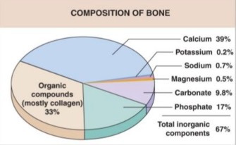
Human bone tissue consists of organic compounds (mostly collagen type-1 proteins), inorganic calcium hydroxyapatite (Ca10 (PO4)6 (OH)2), as well as smaller amounts of potassium, sodium, magnesium, and several other trace minerals.10 It is highly vascularized, metabolically active, and constantly responding to environmental stressors and mechanical stimuli.11,12 (Figure 1)

Figure 1
Figure 1.Composition of Bone

Bone constantly renews itself in a process called remodeling, which is carried out by 2 specialized cell types: osteoclasts and osteoblasts. Osteoclasts remove old, or damaged bone tissue, transferring the tissue components back into the bloodstream. Osteoblasts, under normal physiological conditions, will fill the small holes created by osteoclastic activity with a “mortar” of calcium, minerals, and collagen, forming new bone tissue.

A majority of bone mass is accrued in early adulthood, peaking around the age of 30 years.13 In a healthy adult, the rate of bone formation is roughly equivalent to the rate of bone resorption. However, in osteoporosis, the normal balance of osteoclast and osteoblast activity is altered resulting in accelerated resorption and/or decreased bone formation. As more tissue is resorbed than formed, bones become porous, increasingly fragile and vulnerable to fracture.

Osteoporosis can be classified into 2 distinct types. Primary osteoporosis is associated with normal aging and results from diminishing levels of male and female sex steroid hormones, which degrades the bone trabeculae and decreases mineral content.14,15 Secondary osteoporosis is osteoporosis caused by medical conditions or the use of medications that can interfere with bone reformation.16 Secondary osteoporosis can result from many causes, including disease, specific medication use, nutritional deficiencies, sedentary lifestyle, excessive alcohol consumption, and smoking. (Table 1) However, disorders of calcium metabolism (e.g., hyperparathyroidism, vitamin D deficiency) and bone-demineralizing medications, accounting for approximately 78% of the secondary causes.17

Table 1.Known secondary causes of osteoporosis; congenital and acquired diseases.
Endocrine/Metabolic Hematologic/Gastrointestinal Genetic/ Miscellaneous
Hyperparathyroidism Leukemia Inflammatory arthritides
Hyperthyroidism Multiple myeloma Osteogenesis imperfecta
Hypogonadism Thalassemia Gaucher’s disease
Cushing’s Celiac disease Hypophosphatemia
Diabetes mellitus Gastric bypass surgery Hemochromatosis
Acromegaly Intestinal malabsorption Homocystinuria
Hyperprolactinemia Inflammatory bowel disease Cystic fibrosis
Growth hormone deficit Chronic liver disease Marfan syndrome

Diagnostics

Bone strength – here defined as the ability to withstand trauma without fracturing – is primarily a function of bone density. 75-90% of bone strength is directly related to bone mineral density (BMD).18 While BMD is not the only factor relating to bone strength, previous work has found that BMD is a quantifiable culmination of an individual’s history, genetics, risk factors, and treatment.19

Dual Energy X-ray Absorptiometry

Dual Energy X-ray Absorptiometry (DXA) is the most widely used method for measuring BMD and therefore is a major component of assessing fracture risk.20 Generally, DXA of the lumbar spine and proximal femur (which is subdivided to the femoral neck and total hip) are considered the gold standard measurements for BMD, as they not only quantify the ratio of bone formation to resorption, but also provide a normalized score that can be tracked over time.1,21,22 DXA is a specialized x-ray device that accurately measures bone mineral density; essentially the amount of calcium per area of bone. The results from a DXA test are expressed as T-scores, and are used by the World Health Organization (WHO), as well as the International Osteoporosis Foundation (IOF), to define osteoporosis (see Table 2).23 T-scores are a statistical representation of an individual’s BMD expressed in standard deviations difference from a sample of 20–29-year-old Caucasian females’ femoral neck measurement in the National Health and Nutrition Examination Survey (NHANES) III reference database.24 There is some controversy of these metrics, given the widespread application of female BMD measurements to their male counterparts.25,26 Regardless, the current international standard is to begin treatment (medication) for all patients with a T-score of less than -2.5.27 (Table 2)

Table 2.WHO Categorization and T-Score Limits for Osteopenia and Osteoporosis
WHO category T-score
Normal >-1.0
Osteopenia -1.0 to -2.49
Osteoporosis <-2.5
Established or Severe osteoporosis <-2.5 with one or more fragility fractures

The T-scores above are a normalized comparison to lifetime peak bone mass (30-year-old) of the same gender and ethnic group.

Generally, the accuracy of DXA in calculating BMD is excellent; however, it is important to note that the presence of arthritic changes, especially at the lumbar spine, can falsely elevate resulting T-scores.28 However, DXA results at the lumbar spine can detect early bone loss, in patients over 64 years of age, the proximal femur is more reliable as degenerative changes of the lumbar spine can render the results unusable.29,30 In the event that lumbar and proximal femur DXA examinations are not possible (e.g. immobility, obesity, hip replacements) forearm measurements can also be used.31 Forearm measurements should also be used in the case of hyperparathyroidism, as this location will exhibit changes earlier than the lumbar and proximal femur (CITE).31

The need and/or frequency for repeat DXA tests is situational. Patients at low risk may only need long interval (5-10 years) follow up while patients at high risk and/or newly initiated treatment often are prescribed a 1-year repeat DXA. Intervals between tests are lengthened after a positive response to treatment.32

Other Diagnostics

In addition to DXA results, the fracture risk assessment tool (FRAX) is used to calculate the 10-year probability of fracture. The tool examines age, sex, weight, height, history of previous fracture, parental hip fracture, current smoking, use of glucocorticoids, rheumatoid arthritis, secondary osteoporosis, alcohol (3 or more units/day), and femoral neck BMD.33,34 However, Although the FRAX tool is simple to use, it is an extrapolation and estimation of a future fracture rather than a direct measurement of current bone density as is provided by DXA.

Trabecular bone scores (TBS) have also been used diagnostically. TBS is an indirect indicator of bone microarchitecture and attempts to predict fracture risk by assessing bone quality. Though it is a useful tool, it is recommended as a complement to BMD results, rather than a standalone assessment.35

Peripheral BMD measurements of the tibia and calcaneus are also possible using Quantitative Ultrasound (QUS). QUS devices directly measure the speed of transmission of sound through the bone being assessed and interpolate this to a T-score. These devices have not been found to be reliable for measuring yearly changes in BMD, however - and according to the WHO - QUS should not be used to diagnose osteoporosis.36

Laboratory Testing

If a patient’s DXA scan returns low T-scores, or there is suspicion of conditions affecting the skeleton, laboratory tests should be ordered. Laboratory testing can be thought of as existing in 2 related levels. First level testing can uncover secondary causes of decreased BMD. Indeed, if the first level tests are normal, approximately 90% of secondary osteoporotic causes can be ruled out. On the other hand, an abnormal first level lab warrants further, more specific, laboratory tests and investigation; this is second level testing.37 (Table 3)

Table 3.First and Second Level Laboratory Testing for Osteoporosis
First Level Second Level
Complete blood count (CBC) Serum electrophoresis
Comprehensive metabolic panel Parathyroid hormone (PTH)
25-OH-vitamin D Thyroid stimulating hormone (TSH)
C-reactive protein (CRP)
24-hour urinary calcium
Undercarboxylated Osteocalcin (uOC)

Bone turnover markers are mainly used in clinical trials to monitor efficacy and response to therapy by assessing new bone formation or resorption and/or to evaluate patient adherence to treatments. The tests for new bone formation include osteocalcin, bone isoenzyme of alkaline phosphatase (B-ALP), and type I collagen peptides. The markers for bone resorption are urinary pyridinoline (PYR), urinary deoxypyridinoline (DPYR), and serum levels of type I collagen telopeptides (NTx, CTx).38,39 These are not used, nor recommended, as routine tests in the management of individual patients.

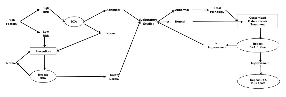
Figure 2 outlines a flow which providers can use to enhance their clinical diagnostics. The decision tree begins in the upper left-hand corner, by assessing a patient for risk factors. High risk individuals are screened using DXA testing. T-scores falling into pathological ranges can then be investigated with first and potentially second level laboratory tests. The information gathered from the DXA, and laboratory tests can then be used to tailor a personalized treatment program.

Figure 2
Figure 2.Osteoporosis Decision Tree

Risk Factors

Risk factors for osteoporosis can be categorized as either modifiable or non-modifiable.40–42 Modifiable factors are those that can be affected through lifestyle choices, therapeutic interventions and/or behavior. On the contrary, non-modifiable factors are mainly genetic or physiologic and are thus more difficult to affect. It is also important to note that risk factors, though important for clinical decision-making, do not accurately predict bone density.43

Modifiable Risk Factors

Medication and Drug Usage

There are a number of medications that can facilitate bone wasting (see Table 4), though it is important to note that the extent and severity of bone loss from these medications is dependent on dose and duration.44 However, of these medications, long term corticosteroid therapy is most commonly associated with drug-induced osteoporosis with the most rapid declines of bone mineral in the first 6 months of use.45,46

Table 4.List of Known Bone Wasting Medications
(Ranked in order of risk*)
Corticosteroids
Aromatase inhibitors
GnRH inhibitors / hormone blockers
Progesterone-only contraception (injectable, implant, highest dose IUD)
Anticonvulsants
Antiretroviral drugs
Vitamin K-antagonist anticoagulant
Statins
Inhaled corticosteroids
Methotrexate
SSRI
Antibiotics (prolonged, oral)
Protein pump inhibitors

*The extent of bone wasting is dose and duration dependent.

Progesterone-only birth control methods can also interfere in bone formation and resorption. Injectable Depo-Provera has been found to cause significant bone demineralization.47,48 Remineralization often occurs with cessation, however this is impacted by duration and age of use.49,50 Women closer to menopause are less able to rebuild bone density. Lower dose progesterone only contraceptives and combined estrogen-progesterone contraceptives have generally not been shown to suppress bone health.51

Diet

The consumption of high and ultra-processed foods and refined sugars, as well as inadequate protein and nutrient intake have been linked to osteoporosis. Ultra-processed foods are lacking in key nutrients required for bone health and often contain high-fructose corn syrup, hydrogenated oils, hydrolyzed proteins, or additives designed to increase flavor (such as flavor enhancers, colors, emulsifiers, emulsifying salts, sweeteners, and thickeners).52

In particular, these foods do not have adequate protein, fiber, vitamins A, C, D, and E, zinc, potassium, phosphorus, magnesium, and calcium, needed to support healthy bone.53 Similarly, the habitual consumption of high sugar drinks, such as soda, has been shown to be a risk factor for osteoporotic fractures.54,55 Sugar increases the urinary excretion of calcium, increasing inflammation, and hyperinsulinemia, hastening bone resorption.56

Adequate dietary protein is also essential for bone health. Approximately 50% of bone volume and 33% of bone mass is composed of structural proteins57; proteins are also involved in the secretion of bone-forming hormones, bone-related nutrient absorption and calcitriol synthesis.58

Inadequate nutrient intake has also been linked to osteoporosis and skeletal health. Vitamins B6, B9 (folic acid), B12, C, D, K are important for bone health, and deficiencies can lead not only osteopenia and osteoporosis, but also to disease states such as rickets and osteomalacia.59–61 Minerals such as calcium, boron, magnesium, manganese, strontium, zinc, and copper are also necessary for optimal bone health.62–67

Excessive alcohol consumption has direct and indirect effects on bone. Excessive is defined as more than two drinks per day for men and more than one drink per day for women.68 Alcohol often displaces good nutrition, has a toxic effect on osteoblasts, and increases the risk of falls. In contrast, modest consumption of alcohol has minimal or even a protective effect.68

Sedentary Lifestyle

Chronic unloading and/or weightlessness can lead to a rapid decline in bone density.69 Sedentary individuals are at risk of lower BMD due to the lack of mechanical stress placed on the bones.70,71 In the extreme, osteosarcopenia – the combination of bone and muscle wasting that can result from inactivity – increases the risk for both falls and fractures.72

Smoking

Smokers have twice the risk of osteoporotic fractures as non-smokers.73 Smokers have approximately 10% lower circulating levels of 1,25-dihydroxyvitamin D, the activated form of vitamin D.74,75 In a study of identical twins, where only 1 of twins smoked, bone mineral was approximately 10% lower. This is leading evidence that smoking is an independent risk factor for osteoporosis.76

Environment

Vitamin D deficiency is a global problem with approximately 42% of the US population being deficient.77 Sunshine (specifically ultraviolet B) striking exposed skin is the main source of vitamin D.78

For the same amount and time in sunshine, darker skinned individuals will absorb less UV radiation.79 The reason for this is the relative differences in skin melanin concentrations. Indeed, vitamin D deficiencies are more common in dark-skinned individuals.80,81 Studies investigating this phenomenon have found as many as 82% of African Americans suffering from hypovitaminosis D, followed closely by Hispanics at 70%.82

Certain environmental substances may increase osteoporosis risk. There is a possible link between osteoporosis and heavy metal exposure, such as cadmium (Cd) and lead (Pb), and industrial chemicals such as phthalates and per- and poly-fluoroalkyl substances (PFASs).83,84 However, these findings have been found in observational studies only and require further exploration.

Non-Modifiable Factors

Ethnicity

Bone density also varies among different ethnicities. Previous studies have found that Africans have higher BMD and lower fracture rates as compared with both Asians and Caucasians. Asians have an approximately 50% lower risk of a hip fracture than Caucasians.85,86

Genetics

Genetics have been found to account for 40-60% of peak bone mass. As greater lifetime peaks of bone density have been associated with lower risks of fracture over time, genetic influences play a significant role in the risk of osteoporosis and fracture.87–89

Age

For a given bone mineral density, fracture risk increases as bone quantity and quality deteriorates with age. In addition, decreases in muscle mass, strength, and balance (common issues associated with age) all increase fall and fracture risk.90,91 It is also important to note that a history of previous fragility fracture – a fracture from low energy trauma – is an independent risk factor for subsequent fractures and increasingly common with and indicative of osteoporosis.

Sex and Hormones

The single greatest contributor to bone mineral loss and fracture risk is decreased hormone levels. In women, the abrupt reduction in estrogen that occurs with menopause leads to increased osteoclastic and decreased osteoblastic activity, resulting in an acceleration of bone mineral losses.92,93

In early stages, this bone loss is found primarily in trabecular bone. Eventually, bone losses will be found in both trabecular and cortical bone.93 In the 4-8 years transition period to menopause, average BMD loss is 10%.93 Nearly half of women lose bone even more rapidly than this, with losses increasing up to 20%.94 This rate of loss can be compounded by earlier onset of menopause; the earlier the onset, the higher the risk of greater bone loss.95

In men, osteoporosis is underestimated and undertreated. Male fracture risk from osteoporosis is also related to decreasing hormone levels, though it presents with a different pattern of bone loss.96 Trabecular bone in men does tend to become thinner, but it maintains its connectivity better than in females.97 Fractures in men also occur 5-7 years later in life as compared with women98 and are often driven by secondary osteoporosis at approximately twice the rate of women.99,100 The risk of hip fracture in elderly men is 5-6% as compared with 16-18% for women. This translates to approximately 30% of hip fractures are being experienced by men.101

Prevention and Treatment

Diet

Dietary guidelines for the prevention of osteoporosis include eating whole foods, consuming adequate protein, having a diet with many dark green leafy vegetables, as well as avoiding excess sugar, alcohol, and smoking.102 Individuals should avoid heavily processed foods, and ensure they are achieving adequate vitamin and mineral intake from their diet.103 Recommended protein intake is 1.0-1.2 g/kg body weight/day.104

Calcium is undeniably important for bone health. The recommended daily allowance (RDA) for calcium for adults is 1000-1200 mg/day, though some studies have disputed this amount, suggesting instead a practical allowance of 400-500 mg/day.105 Several studies have even found that less than 300 mg/day will still confer benefits.106,107 Good dietary sources of calcium include kale, sardines, milk and cook beans, and can provide 100-350 mg/serving (e.g. 1 cup cooked collards or kale, 3 ounces of sardines, 8 ounces of milk, or 4 ounces of cooked beans). Many foods can supply or surpass the RDA (see Appendix 1).

Overconsumption of calcium does not appear to add any measurable benefit; however, with deficiency, supplementation is advantageous.108,109 Previous literature has found a strong correlation between calcium, dairy consumption, and bone health.110,111 Interestingly, though calcium is undeniably a component of bone, numerous trials have found little or no correlation between calcium (or dairy) consumption and bone mineral density and/or rate of fractures.108,112–114

Calcium is not always readily available in every diet and, as such, supplementation is advised. There are numerous, conflicting, opinions as to the preferred form of calcium supplementation. Considerations must include solubility, molecular size, pill size, particle size per manufacturer, as well as cost. Table 5 displays some of the benefits and drawbacks of four commonly available forms.

Table 5.Four Common Forms of Calcium Supplementation
Calcium form Calcium % Benefit Drawback
Carbonate 40 Least cost per mg. Highest concentration. Must be taken with meal.
Poor absorption in presence of antacids.
Citrate 21 Highly soluble.
May take anytime.
Absorbed even with antacids (H-2 blockers or PPI).
Gluconate 9 Highly soluble Low concentration
Lactate 13 Highly soluble Low concentration

Calcium citrate appears to be the best choice when considering solubility, versatility, and cost. There are other forms of calcium supplements beyond the four listed above; hydroxyapatite, citrate-malate, acetate. None of these seem to have any advantage over citrate.115

Beyond bioavailability, calcium absorption is itself multifactorial. Optimal levels of Vitamin D can facilitate greater calcium absorption, as well as taking calcium with other foods and in smaller doses over a greater number of meals. Pregnancy has also been found to increase calcium absorption rates. Conversely, foods containing oxalates and/or phytates, low serum vitamin D levels and hypo or achlorhydria can decrease calcium absorption.116

Though the RDA for vitamin D is 400-800 IU/day, there is growing evidence that this may not be enough.117–121 Levels of Vitamin D are quantified by measuring blood concentrations of 25,OH-vitamin D (see Table 6).

Table 6.Vitamin D Concentrations for Normal, Insufficient and Deficient
Normal >30 ng/ml (nanogram per milliliter)
Insufficient 20-30 ng/ml
Deficient <20 ng/ml

It is important to note, however, that calcium and vitamin D have both received disproportionate attention when discussing diet. While both compounds contribute to skeletal health, neither is enough to address the wide variety of components necessary to build and maintain bone.1,122 Vitamins C and K, as well as B6, B9 and B12 are also important for bone health.123–125

Vitamin C appears to influence bone health in several ways. Vitamin C suppresses osteoclast activity, acts as a co-factor for osteoblast differentiation, enhances intestinal calcium absorption, and is needed for collagen formation.126 Adequate intake is positively associated with higher BMD127 as well as lower rates of hip fracture.128 Vitamin C can be found in fresh produce (see Appendix).

Vitamin K, especially K2M7, has been shown to have a synergistic effect with vitamin D, further promoting bone health.129,130 Vitamin K is multifunctional and has been demonstrated to promote bone formation by stimulating osteoblast differentiation and inhibiting osteoblast apoptosis,131 regulating extracellular matrix mineralization,132 and decreasing bone resorption by inhibiting osteoclast differentiation.131 Vitamin K has also been shown to induce and activate osteocalcin (bone GLA protein),133–135 a compound that binds calcium ions to hydroxyapatite crystals.132,136 Providers should also be aware that the use of statin drugs can inhibit the formation of vitamin K-dependent proteins, specifically the activated form of osteocalcin; carboxylated osteocalcin (cOC).137 Clinical trials using vitamin K in isolation yielded small, statistically insignificant, increases in bone mineral and minimal fracture reduction.138,139 However, combining vitamins D and K [as K2M4140 and K2M7141] yield positive, statistically significant improvements.

The RDA for magnesium is 310-360 mg and 400-420 mg for women and men, respectively.142 Approximately 50–60% of the total body magnesium content is accumulated in bone, and magnesium deficiency has been associated with reduced bone formation.143–145 Magnesium is also a co-factor needed for the activation of vitamin D.146 However, though there is leading evidence for an important role for magnesium in osteoporosis, no randomized studies have evaluated the effects of magnesium in isolation, and there is only a weak association between low serum magnesium levels and osteoporosis.147,148

Several trials demonstrate the benefits of trace minerals including magnesium, manganese, zinc, boron, and strontium.149–155 Table 7 provides RDA values for these minerals.

Table 7.USRDA values for Assorted Minerals
USRDA for adults; mg/day
Magnesium 310-420
Manganese 2.3-2.6
Zinc 8-13
Boron N/A
Strontium N/A

Compounds other than protein, vitamins and minerals have also received research attention. Collagen, Cissus quadrangularis, soy isoflavones, ipriflavone, DHEA, melatonin, and dried plums (prunes) have all been examined. Small and/or short duration studies looking at the effects of collagen156,157 and Cissus quadrangularis158 show encouraging results, though longer-term studies are needed.

In contrast, several well-done studies using soy isoflavones, ipriflavone, DHEA, and dried plums have demonstrated consistent benefit for bone health.159–162 Therapeutic doses of both soy isoflavones and ipriflavone should be restricted to use in women due to their estrogenic properties. DHEA is an adrenal hormonal which can be converted into a number of sex steroid hormones. Though it has inherent potency, more research is needed to understand all of its effects at varied doses. Dried plums have been subjected to several trials with sound methodology, yielding positive results. Studies have found leading evidence that consumption can increase bone formation, increase trabecular microstructure, decrease osteoclastogenesis, and decrease RANKL (a compound leading to bone resorption).163–168

Melatonin appears to both enhance anabolic effects and have antiresorptive benefits. Melatonin improves bone formation by promoting the differentiation of mesenchymal cells into osteoblasts. Bone resorption is reduced by diminishing the synthesis of RANKL.169,170

Combinations of micronutrients also appear to offer greater benefit than single nutrient treatments for osteoporosis. The Combination of Micronutrients for Bone Study (COMB) investigated 77 osteoporotic patients who either declined or failed with a trial of pharmacotherapy. These patients were offered a nutrient combination that included vitamin D3, vitamin K2, strontium, magnesium, and fish oil derived DHA (docosahexaenoic acid). After 12 months, the repeat DXA demonstrated statistically significant improvement of BMD at the spine, hip, and femoral neck sites. There were no fractures in the treatment group.171

The Melatonin-micronutrients Osteopenia Treatment Study (MOTS) was a 1-year double-blind randomized controlled trial using a combination of melatonin, strontium, vitamin D3, vitamin K2 (MK-7) that measured bone mineral, bone markers, and quality of life for postmenopausal osteopenic women. The treatment group had significant increases in BMD which were mirrored by changes in numerous markers of increased bone formation and decreased bone resorption. The intended improvements in skeletal health were accomplished in addition to benefits in mood and sleep quality. This well-designed combination of micronutrients appears to offer an effective treatment option and can be safely used with osteopenic patients, prior to further declines in BMD.172

Neither the COMB nor MOTS study described any adverse events or unintended consequences during the one-year duration of the trials. Though safety beyond one year duration is unclear, these nutrient combinations appear to be safe and effective within that time frame. Not all combination supplement trials show benefit.173

Medications (Table 8)

The list of prescription treatments has grown substantially since 1995, when the first osteoporosis-specific medications were approved by the United States Food and Drug Administration (USFDA). These medications can be categorized either by hormone vs. non-hormone, antiresorptive vs. anabolic, oral vs. injection, or first line vs. second line.

Table 8.List of Common Osteoporosis Medications
Class and drug Brand name Form Frequency Antiresorptive vs. anabolic Hormone vs. non-hormone 1st vs. 2nd line
Bisphosphonates
Alendronate Fosamax Oral Daily, weekly Antiresorptive Nonhormone 1st
Alendronate Binosto Oral Weekly Antiresorptive Nonhormone 1st
Ibandronate Boniva Oral Monthly Antiresorptive Nonhormone 1st
Ibandronate Boniva IV Quarterly Antiresorptive Nonhormone 2nd
Risedronate Actonel Oral Daily, weekly Antiresorptive Nonhormone 1st
Risedronate Atelvia Oral Weekly Antiresorptive Nonhormone 1st
Zoledronic acid Reclast IV Annual Antiresorptive Nonhormone 2nd
RANKL inhibitor
Denosumab Prolia Injection 2x/annual Antiresorptive Nonhormone 1st, 2nd
Calcitonin
Calcitonin Fortical, Miacalcin Intranasal Daily Antiresorptive Hormone 1st
Miacalcin Injection Variable Antiresorptive Hormone 1st
Estrogen
Estrogen Multiple products Oral Daily Antiresorptive Hormone 1st
Multiple products Trans-dermal 2x/week Antiresorptive Hormone 1st
SERMs
Raloxifene Evista Oral Daily Antiresorptive Hormone 1st
Estrogen-SERM combination
Estrogen-bazodoxifene Duavee Oral Daily Antiresorptive Hormone 2nd
Sclerostin inhibitor
Romosuzumab-aqqg Evenity Injection 1x/month/ 12 months Anabolic Hormone 2nd
Parathyroid hormone analog
Teriparatide Forteo Injection Daily/18 months Anabolic Hormone 2nd
Parathyroid hormone-related protein analog Tymlos Injection Daily/18 months Anabolic Hormone 2nd

Each of the above medications has its own share of risks and benefits and all confer varying degrees of fracture prevention. The risk and side effects are seen in table 9.

Table 9.Risks and side effects of medications.
Drug or drug class Minor side effects Serious side effects
Oral bisphosphonates GI irritation, gastric reflux, constipation Atypical femur fracture, osteonecrosis of the jaw, esophageal ulcer
IV bisphosphonates Self-limiting mild fever Atypical femur fracture, osteonecrosis of the jaw
Denosumab Weakness
Muscle, spine, extremity pain
Increase susceptibility to infections
Intranasal calcitonin Nasal irritation, nose bleed, headache
Estrogen, oral Bloating, nausea, vaginal bleeding, breast tenderness/swelling, and fluid retention Blood clots, Stroke, Heart attack
Breast, uterine, ovarian cancers
Estrogen, transdermal Bloating, headache, breast tenderness Blood clots, Stroke, Heart attack
Breast, uterine, ovarian cancers
SERMs Hot flashes, GI upset, joint pain Blood clots
Stroke
Estrogen-SERM Diarrhea, GI upset, water retention Blood clots, Stroke, Heart attack
Breast, uterine, ovarian cancers
Sclerostin inhibitor Joint pain, headache, insomnia Heart attack, stroke
Atypical femur fracture
Osteonecrosis of the jaw
Parathyroid hormone analog Joint pain, headache, nausea Osteosarcoma
Seizures

However, compliance with these medications can be poor, with 20-30% of patients stopping treatment within 6-12 months.174,175 Reasons for non-compliance include both the actual side effects as well as the fear of reported risk.174

Exercise

Exercise-based therapies should seek to safely increase skeletal loading and stimulate positive osseus adaptations to mechanical stress. The overall goal of training should be to reduce the risk of falls and improve strength and mobility.176 Walking has been found to increase muscle tone and overall fitness, as well as reduce the number of fall events, though only shown minimal changes in BMD.177 Resistance and higher impact training paradigms such as weightlifting and running offer greater benefits to BMD, while also facilitating strength and balance.178,179

Yoga,180 tai chi,181 unipedal standing,182 and walking,177 though less beneficial for BMD than impact exercise, have shown significant value for overall health, balance, and fracture reduction.

Some studies have suggested that vibration of the whole body or regional areas offers potential benefit for bone formation; however, these studies have differing protocols, used various endpoints, and were relatively short term.183,184 As such, the parameters of the therapy and results of care have been inconsistent.

Summary of Recommendations

Numerous professional groups have put forth guidelines germane to the diagnosis, prevention and treatment of osteoporosis. Guidelines from the American Association of Clinical Endocrinologists, American College of Endocrinology, North American Menopause Society, Endocrine Society, American College of Rheumatology, National Osteoporosis Foundation, and the American College of Physicians are all relatively consistent and focus almost entirely on treatment with medication.185

It is important to note that a single treatment for all osteoporotic patients is unrealistic given varied etiologies. Potential treatments and preventive protocols should be seen as guidelines that require personalization. The intervention for a 16-year-old with anorexia, a 25-year-old elite athlete or an 80-year-old senior cannot be identical. The rate of BMD decline is variable, and is influenced by genetics, hormonal status, nutrition, exercise, some disease processes, and medications.45 Treatments can – and should – be as numerous and comprehensive as the number of risk factors for this disease and take sex, activity level, lifestyle and comorbidities – at a minimum – into account.

Gold standard testing and diagnosis currently utilizes guidelines established by the WHO based on DXA testing. T-scores from DXA testing provide adequate summation of the spectrum of factors that can influence bone health (e.g. genetic, lifestyle, medications, exercise, etc.), but does not specify the underlying reason(s). Additionally, examination of T-scores over time provide a means to evaluate trending and responses to treatment. Follow-up DXA testing should be as dynamic as skeletal health and based on an individual’s unique presentation. In an older adult with a T-score necessitating treatment, follow up in 1-year is warranted. If the follow up DXA demonstrates an increase in BMD after the initiation of treatment, then greater intervals between tests is appropriate. In a patient with low risk and a normal DXA result, a repeat test in 5-10 years is reasonable. Providers should note that there are known shortcomings with these criteria – specifically that the normative values T-scores are based on rely entirely on a sample of under 30-year-old Caucasian women24 and thus may be less accurate when approximating BMD in men, as well as both sexes of other ethnicities. Laboratory tests can also be helpful in identifying secondary causes of osteoporosis (such as hyperparathyroidism, vitamin D deficiency or kidney disease) following an unexpectedly low DXA test. If laboratory tests are abnormal, further testing is often indicated. A battery of both DXA and laboratory examinations can be used to create an individualized treatment program.

Prevention and treatment strategies for osteoporosis should begin with a balanced nutritious diet. General dietary guidelines for the prevention of osteoporosis include eating whole foods, consuming adequate protein, having a diet with many dark green leafy vegetables, as well as avoiding excess sugar, alcohol, and smoking.186 Individuals should avoid heavily processed foods, and ensure they are achieving adequate vitamin and mineral intake from their diet.103 Though calcium and vitamin D are important to bone health, they are far from the only vitamins and minerals needed. Supplements should be considered in the event that diet alone cannot supply all of the needed bone building nutrients. It is also important to note that vitamins and minerals do not operate independently; rather, the absorption and utilization of one may rely entirely on the presence of another. Dosage, schedule of intake and bioavailability are all important factors to consider in designing a nutritional intervention. (Table 10).

Table 10.Lifestyle Behaviors and Supplements Known to Enhance Bone Health
Category Factors Source
Balanced diet Protein; 1.0 – 1.2 g/kg body weight

Calcium

Minerals
Meat, legumes, dairy, nuts

Dairy, broccoli family, calcium-fortified foods

Dark green leafy vegetables, organ meat, most vegetables
Exercise High impact
Balance
Running, jumping, volleyball, weight lifting.
Tai chi, unipedal standing, yoga.
Vitamin D3 Exposure to noon-time sunshine or
1000-5000 IU/day
Blood level should be a minimum of 30 ng/ml

Supplements to be taken in morning.
Vitamin K2 100-500 mcg/day Contraindicated if patient is taking a vitamin K-antagonist anticoagulant.
Magnesium 250 mg/ day Found in greens, nuts, seeds, beans, whole grains
Melatonin 5 mg Take at bed time
Strontium 500-800 mg/ day Found in bran, root vegetable peels, spinach, lettuce.
Wild fish oil 250 mg/day Docosahexanoic acid (DHA), Eicosapentanoic acid (EPA)
Calcium RDA is 800-1000 mg/day From food and/or supplements.
Dried plums (prunes) 50 grams or 5-6 prunes/ day Bowel tolerance increases with unsulfured prunes.
Vitamin C 500-1000 mg/ day Found in raw red peppers, citrus, kiwi
Boron 2 mg/ day Found in coffee, milk, apples, potato, beans
Manganese 10-20 mg/ day Found in shellfish, brown rice, hazelnuts, black tea
Zinc 10-20 mg/ day Found in meat, shellfish, legumes, seeds

Nutrient interventions can be categorized into diet, supplementation to insure quantities consistent with the USRDA, and pharmacologic doses. Diet sources and RDA quantities are associated with minimal risk. The risk: benefit relationship must be considered when prescribing any mega or pharmacologic doses or combinations. Additionally, nutrient-drug interactions may also impart risk or additional benefits.

Table 10 is a composite of lifestyle and supplements shown to enhance bone health. To date, there is no single study using the entirety of the summary table. Rather, this list expands upon the successful COMB and MOTS trials.171,172

Exercise based on increased skeletal loading and impact can help to build and maintain bone. Exercises such as volleyball, dancing, and weight training are all helpful for bone formation.187 Walking and swimming are excellent cardiovascular exercises, but less helpful for creating the forces necessary to stimulate bone formation. There is indirect evidence for yoga and tai chi in maintaining muscle mass and balance as well as reducing fall risk.

CONCLUSION

The literature is limited on the subject of manual therapy risk or benefit for osteoporotic patients. However, in regard to safety, one review concluded that Grade II to IV mobilization techniques were used with no serious adverse events reported.188 Providers should use best judgment with treatment recommendations.

The adaptive process of assessing risk, DXA and laboratory testing, prescribing dietary/supplement/exercise/lifestyle interventions that are customized to individual patient’s needs requires more provider effort, but in the end will yield more positive outcomes than management limited to medications or single nutrients.

References

1.
Cosman F, de Beur SJ, LeBoff MS, et al. Clinician’s guide to prevention and treatment of osteoporosis. Osteoporos Int. 2014;25(10):2359-2381. doi:10.1007/s00198-014-2794-2
Google ScholarPubMed CentralPubMed
2.
Sözen T, Özışık L, Başaran NÇ. An overview and management of osteoporosis. Eur J Rheumatol. 2017;4(1):46-56. doi:10.5152/eurjrheum.2016.048
Google ScholarPubMed CentralPubMed
3.
Hansen D, Bazell C, Pelizzari P, Pyenson B. Medicare cost of osteoporotic fractures: The clinical and cost burden of an important consequence of osteoporosis. Published online August 2019. Accessed January 21, 2021. https://static1.squarespace.com/static/5c0860aff793924efe2230f3/t/5d76b949deb7e9086ee3d7dd/1568061771769/Medicare+Cost+of+Osteoporotic+Fractures+20190827.pdf
4.
Fractured neck of femur. Prevention and management. Summary and recommendations of a report of the Royal College of Physicians. J R Coll Physicians Lond. 1989;23(1):8-12.
Google Scholar
5.
Cooper C, Campion G, Melton LR III. Hip fractures in the elderly: a world-wide projection. Osteoporos Int. 1992;2(6):285-289. doi:10.1007/bf01623184
Google Scholar
6.
Prentice RL, Pettinger MB, Jackson RD, et al. Health risks and benefits from calcium and vitamin D supplementation: Women’s Health Initiative clinical trial and cohort study. Osteoporos Int. 2013;24(2):567-580. doi:10.1007/s00198-012-2224-2
Google ScholarPubMed CentralPubMed
7.
Department of Health and Human Services. Bone Health and Osteoporosis: A Report of the Surgeon General. Department of Health and Human Services, Office of the Surgeon General; 2004.
Google Scholar
8.
National Institutes of Health Office of Medical Applications of Research. Osteoporosis prevention, diagnosis, and therapy. NIH Consensus Statement. 2000;17(1):1-45.
Google Scholar
9.
World Health Organization. WHO Scientific Group on the Assessment of Osteoporosis at Primary Health Care Level: summary meeting report. Published online May 5–7, 2004. Accessed December 27, 2017. https://www.who.int/chp/topics/Osteoporosis.pdf
10.
Baron R. In: Favus MJ, ed. Primer on the Metabolic Bone Diseases and Disorders of Mineral Metabolism. ASBMR; 2003:1-8.
Google Scholar
11.
Senderovich H, Kosmopoulos A. An Insight into the effect of exercises on the prevention of osteoporosis and associated fractures in high-risk individuals. Rambam Maimonides Med J. 2018;9(1):e0005. doi:10.5041/rmmj.10325
Google ScholarPubMed CentralPubMed
12.
Frost HM. A 2003 Update of bone physiology and Wolff’s Law for clinicians. Angle Orthod. 2004;74(1):3-15. doi:10.1043/0003-3219(2004)074<0003:AUOBPA>2.0.CO;2
Google Scholar
13.
Baxter-Jones ADG, Faulkner RA, Forwood MR, Mirwald RL, Bailey DA. Bone mineral accrual from 8 to 30 years of age: An estimation of peak bone mass. J Bone Mineral Res. 2011;26(8):1729-1739. doi:10.1002/jbmr.412
Google Scholar
14.
Raisz LG. Pathogenesis of osteoporosis: concepts, conflicts, and prospects. Journal Clin Invest. 2005;115(12):3318-3325. doi:10.1172/jci27071
Google ScholarPubMed CentralPubMed
15.
Hunter DJ, Sambrook PN. Bone loss: epidemiology of bone loss. Arthritis Res. 2000;2(6):441-445. doi:10.1186/ar125
Google ScholarPubMed CentralPubMed
16.
Nuti R, Brandi ML, Chechia G, et al. Guidelines for the management of osteoporosis and fragility fractures. Intern Emerg Med. 2019;14(1):85-102. doi:10.1007/s11739-018-1874-2
Google ScholarPubMed CentralPubMed
17.
Tannenbaum C, Clark J, Schwartzman K, et al. Yield of laboratory testing to identify secondary contributors to osteoporosis in otherwise healthy women. J Clin Endo Metab. 2002;87(10):4431-4437. doi:10.1210/jc.2002-020275
Google Scholar
18.
Holroyd C, Crozier S, Inskip H, et al. Mother-child are of greater magnitude that father-child DXA bone associations: Results from the Southhampton women’s survey. Osteoporos Int. 2018;29:S450-S450.
Google Scholar
19.
Pouresmaeili F, Kamalidehghan B, Kamarehei M, Goh YM. A comprehensive overview on osteoporosis and its risk factors. Ther Clin Risk Manag. 2018;14:2029-2049. doi:10.2147/tcrm.s138000
Google ScholarPubMed CentralPubMed
20.
Adams JE. Quantitative computed tomography. European J Radiol. 2009;71(3):415-424. doi:10.1016/j.ejrad.2009.04.074
Google Scholar
21.
Binkley N, Adler R, Bilezikian JP. Osteoporosis diagnosis in men: The T-score controversy revisited. Curr Osteoporos Rep. 2014;12(4):403-409. doi:10.1007/s11914-014-0242-z
Google ScholarPubMed CentralPubMed
22.
Miller PD, Bonnick SL, Rosen CJ. Consensus of an international panel on the clinical utility of bone mass measurements in the detection of low bone mass in the adult population. Calcif Tissue Int. 1996;58(4):207-214. doi:10.1007/bf02508636
Google Scholar
23.
Camacho PM, Petak SM, Binkley N, et al. American Association of Clinical Endocrinologists and American College of Endocrinology: clinical practice guidelines for the diagnosis and treatment of postmenopausal osteoporosis—2016. Endocr Pract. 2016;22(suppl 4):S1-S42.
Google Scholar
24.
Qaseem A, Forciea MA, McLean RM, Denberg TD. Treatment of low bone density or osteoporosis to prevent fractures in men and women: a clinical practice guideline update from the American College of Physicians. Ann Intern Med. 2017;166(11):1-27. doi:10.7326/m15-1361
Google Scholar
25.
Lohman M, Tallroth K, Kettunen JA, Marttinen MT. Reproducibility of dual-energy x-ray absorptiometry and regional body composition measurements using different scanning positions and definitions of regions. Metabolism. 2009;58(11):1663-1668. doi:10.1016/j.metabol.2009.05.023
Google Scholar
26.
Kanis JA, Adachi JD, Cooper C, et al. Standardising the descriptive epidemiology of osteoporosis: recommendations from the epidemiology and quality of life working group of IOF. Osteoporos Int. 2013;24(11):2763-2764. doi:10.1007/s00198-013-2413-7
Google ScholarPubMed CentralPubMed
27.
Kanis JA, Bianchi G, Bilezikian JP, et al. Towards a diagnostic and therapeutic consensus in male osteoporosis. Osteoporos Int. 2011;22(11):2789-2798. doi:10.1007/s00198-011-1632-z
Google ScholarPubMed CentralPubMed
28.
Alsop C. Techniques of bone mass measurement. Endocrine Abstract. 2005;9:s50.
Google Scholar
29.
Mazess R, Collick B, Trempe J, Barden H, Hanson J. Performance evaluation of a dual-energy X-ray bone densitometer. Calcif Tissue Int. 1989;44(3):228-232. doi:10.1007/bf02556569
Google Scholar
30.
Gillette-Guyonnet S, Andrieu S, Nourhashemi F, et al. Comparison of bone mineral density and body composition measurements in women obtained from two DXA instruments. Mech Ageing Dev. 2003;124(3):317-321. doi:10.1016/s0047-6374(02)00199-9
Google Scholar
31.
Wood K, Dhital S, Chen H, Sippel RS. What is the utility of distal forearm DXA in primary hyperparathyroidism? Oncologist. 2012;17(3):322-325. doi:10.1634/theoncologist.2011-0285
Google ScholarPubMed CentralPubMed
32.
White VanGompel EC, Franks P, Robbins JA, Fenton JJ. Incidence and predictors of repeat bone mineral densitometry: A longitudinal cohort study. J Gen Intern Med. 2017;32(10):1090-1096. doi:10.1007/s11606-017-4094-y
Google ScholarPubMed CentralPubMed
33.
Kanis JA, Harvey NC, Johansson H, et al. A decade of FRAX: how has it changed the management of osteoporosis? Aging Clin Exp Res. 2020;32(2):187-196. doi:10.1007/s40520-019-01432-y
Google Scholar
34.
El Miedany Y. FRAX: re-adjust or re-think. Arch Osteoporos. 2020;15(1):150. doi:10.1007/s11657-020-00827-z
Google ScholarPubMed CentralPubMed
35.
Rajan R, Cherian KE, Kapoor N, Paul TV. Trabecular bone score—An emerging tool in the management of osteoporosis. Indian J Endocr Metab. 2020;24(3):237. doi:10.4103/ijem.ijem_147_20
Google ScholarPubMed CentralPubMed
36.
Maggi S, Noale M, Giannini S, et al. Quantitative heel ultrasound in a population-based study in Italy and its relationship with fracture history; the ESOPO Study. Ost Int. 2006;17(2):237-244. doi:10.1007/s00198-005-1985-2
Google Scholar
37.
Crandall C. Laboratory workup for osteoporosis- Which test are most cost-effective. Postgrad Med. 2003;114(3):35-44. doi:10.3810/pgm.2003.09.1492
Google Scholar
38.
Hong L, Liu D, Wu F, Wang M, Cen Y, Ma L. Correlation between bone turnover markers and bone mineral density in patients undergoing long-term anti-osteoporosis treatment: A systematic review and meta-analysis. Appl Sci. 2020;10(3):832-848. doi:10.3390/app10030832
Google Scholar
39.
Zhang T, Liu P, Zhang Y, et al. Combining information from multiple bone turnover markers as diagnostic indices for osteoporosis using support vector machines. J Biomark. 2019;24(2):120-126. doi:10.1080/1354750x.2018.1539767
Google Scholar
40.
Management of osteoporosis in postmenopausal women: 2010 position statement of The North American Menopause Society. Menopause. 2010;17(1):25-54. doi:10.1097/gme.0b013e3181c617e6
Google Scholar
41.
Kawamata A, Iihara M, Okamoto T, Obara T. Bone mineral density before and after surgical cure of Cushing’s syndrome due to adrenocortical adenoma: prospective study. World J Surg. 2008;32(5):890-896. doi:10.1007/s00268-007-9394-7
Google Scholar
42.
Sutton RAL, Dian L, Guy P. Osteoporosis in men: an underrecognized and undertreated problem. BCMJ. 2011;53(10):535-540.
Google Scholar
43.
Stevenson JC, Lees B, Devenport M, Cust MP, Ganger KF. Determinants of bone density in normal women: risk factors for future osteoporosis? Br Med J. 1989;298(6678):924-928. doi:10.1136/bmj.298.6678.924
Google ScholarPubMed CentralPubMed
44.
Panday K, Gona A, Humphrey MB. Medication-induced osteoporosis: screening and treatment strategies. Ther Adv Musculoskelet Dis. 2014;6(5):185-202. doi:10.1177/1759720x14546350
Google ScholarPubMed CentralPubMed
45.
NIH Osteoporosis and Related Bone Diseases National Resource Center. The Surgeon General's report on bone health and osteoporosis: what it means to you. Published online December 2015. Accessed June 6, 2017. https://www.niams.nih.gov/health-topics/bone-health-and-osteoporosis
46.
Buckley L, Guyatt G, Fink HA, et al. 2017 American College of Rheumatology guideline for the prevention and treatment of glucocorticoid-induced osteoporosis. Arthritis Care Res (Hoboken). 2017;69(8):1095-1110. doi:10.1002/acr.23279
Google Scholar
47.
Wong AYK, Tang LCH, Chin RKH. Levonorgestrel-releasing intrauterine system (Mirena®) and depot medroxyprogesterone acetate (Depoprovera) as long-term maintenance therapy for patients with moderate and severe endometriosis: A randomised controlled trial. ANZJOG. 2010;50(3):273-279. doi:10.1111/j.1479-828x.2010.01152.x
Google Scholar
48.
Walsh JS, Eastell R, Peel NFA. Effects of depot medroxyprogesterone acetate on bone density and bone metabolism before and after peak bone mass: A case-control study. J Clin Endocrinol Metab. 2008;93(4):1317-1323. doi:10.1210/jc.2007-2201
Google Scholar
49.
Rosenberg L, Zhang Y, Constant D, et al. Bone status after cessation of use of injectable progestin contraceptives. Contraception. 2007;76(6):425-461. doi:10.1016/j.contraception.2007.08.010
Google Scholar
50.
Banks E, Berrington A, Casabonne D. Overview of the relationship between use of progestogen-only contraceptives and bone mineral density. Br J Obst Gyn. 2001;108(12):1214-1221. doi:10.1016/s0306-5456(01)00296-0
Google Scholar
51.
Hadji P, Colli E, Regidor PA. Bone health in estrogen-free contraception. Osteoporos Int. 2019;30(12):2391-2400. doi:10.1007/s00198-019-05103-6
Google ScholarPubMed CentralPubMed
52.
Monteiro CA, Cannon G, Levy RB, et al. Ultra-processed foods: what they are and how to identify them. Public Health Nutr. 2019;22(5):936-941. doi:10.1017/s1368980018003762
Google ScholarPubMed CentralPubMed
53.
Martínez Steele E, Popkin BM, Swinburn B, Monteiro CA. The share of ultra-processed foods and the overall nutritional quality of diets in the US: evidence from a nationally representative cross-sectional study. Popul Health Metrics. 2017;15(1):6. doi:10.1186/s12963-017-0119-3
Google ScholarPubMed CentralPubMed
54.
Fung TT, Arasaratnam MH, Grodstein F, et al. Soda consumption and risk of hip fractures in postmenopausal women in the Nurses’ Health Study. Am J Clin Nutr. 2014;100(3):953-958. doi:10.3945/ajcn.114.083352
Google ScholarPubMed CentralPubMed
55.
Tucker KL, Morita K, Qiao N, Hannan MT, Cupples LA, Kiel DP. Colas, but not other carbonated beverages, are associated with low bone mineral density in older women: The Framingham Osteoporosis Study. Am J Clin Nutr. 2006;84(4):936-942. doi:10.1093/ajcn/84.4.936
Google Scholar
56.
DiNicolantonio JJ, Mehta V, Zaman SB, O’Keefe JH. Not salt but sugar as aetiological in osteoporosis: A review. Mo Med. 2018;115(3):247-252.
Google Scholar
57.
Boskey AL, Spevak L, Paschalis E, Doty SB, McKee MD. Osteopontin deficiency increases mineral content and mineral crystallinity in mouse bone. Calcif Tissue Int. 2002;71(2):145-154. doi:10.1007/s00223-001-1121-z
Google Scholar
58.
Heaney RP, Layman DK. Amount and type of protein influences bone health. Am J Clin Nutr. 2008;87(5):1567S-1570S. doi:10.1093/ajcn/87.5.1567s
Google Scholar
59.
Herrmann M, Schmidt JP, Umanskaya N, et al. The role of hyperhomocysteinemia as well as folate, vitamin B6 and B12 deficiencies in osteoporosis – a systematic review. Clin Chem Lab Med. 2007;45(12):1621-1632. doi:10.1515/cclm.2007.362
Google Scholar
60.
Leveille SG, LaCroix AZ, Koepsell TD, Beresford SA, Van Belle G, Buchner DM. Dietary vitamin C and bone mineral density in postmenopausal women in Washington State, USA. J Epidemiol Community Health. 1997;51(5):479-485. doi:10.1136/jech.51.5.479
Google ScholarPubMed CentralPubMed
61.
Hall SL, Greendale GA. The relation of dietary vitamin C intake to bone mineral density: results from the PEPI study. Calcif Tissue Int. 1998;63(3):183-189. doi:10.1007/s002239900512
Google Scholar
62.
Pizzorno L. Nothing boring about boron. Integr Med (Encinitas). 2015;14(4):35-48.
Google Scholar
63.
Castiglioni S, Cazzaniga A, Albisetti W, Maier JAM. Magnesium and osteoporosis: Current state of knowledge and future research directions. Nutrients. 2013;5(8):3022-3033. doi:10.3390/nu5083022
Google ScholarPubMed CentralPubMed
64.
Saltman PD, Strause LG. The role of trace minerals in osteoporosis. J Am Col Nutr. 1993;12(4):384-389. doi:10.1080/07315724.1993.10718327
Google Scholar
65.
Reginster JY, Bruyère O, Sawicki A, et al. Long-term treatment of postmenopausal osteoporosis with strontium ranelate: Results at 8 years. Bone. 2009;45(6):1059-1064. doi:10.1016/j.bone.2009.08.004
Google Scholar
66.
Yamaguchi M. Role of nutritional zinc in the prevention of osteoporosis. Mol Cell Biochem. 2010;338(1-2):241-254. doi:10.1007/s11010-009-0358-0
Google Scholar
67.
Lowe NM, Fraser WD, Jackson MJ. Is there a potential therapeutic value of copper and zinc for osteoporosis? Proc Nutr Soc. 2002;61(2):181-185. doi:10.1079/pns2002154
Google Scholar
68.
Turner RT. Skeletal Response to Alcohol. Alcoholism Clin Exp Res. 2006;24(11):1693-1701. doi:10.1111/j.1530-0277.2000.tb01971.x
Google Scholar
69.
Gabel L, Liphardt AM, Hulme PA, et al. Incomplete recovery of bone strength and trabecular microarchitecture at the distal tibia 1 year after return from long duration spaceflight. Sci Rep. 2022;12(1):9446. doi:10.1038/s41598-022-13461-1
Google ScholarPubMed CentralPubMed
70.
Snow CM, Shaw JM, Matkin CC. Physical activity and risk of osteoporosis. In: Marcus R, Feldman D, Kelsey J, eds. Osteoporosis. Academic Press; 1996:511-528.
Google Scholar
71.
Warren MP, Chua AT. Exercise-induced amenorrhea and bone health in the adolescent athlete. Ann NY Acad Sci. 2008;1135(1):244-252. doi:10.1196/annals.1429.025
Google Scholar
72.
Clynes MA, Gregson CL, Bruyère O, Cooper C, Dennison EM. Osteosarcopenia: where osteoporosis and sarcopenia collide. Rheumatology. 2021;60(2):529-537. doi:10.1093/rheumatology/keaa755
Google Scholar
73.
Øyin J, Gjesdal CG, Nyagård OK, Lie SA, Mayer HE, et al. Correction: Smoking and Body Fat Mass in Relation to Bone Mineral Density and Hip Fractures: The Hordaland Health Study. PLoS ONE. 2014;9(6):e101335. doi:10.1371/journal.pone.0101335
Google ScholarPubMed Central
74.
Bijelic R, Milicevic S, Balaban J. Risk Factors for Osteoporosis in Postmenopausal Women. Med Arch. 2017;1(1):25-28. doi:10.5455/medarh.2017.71.25-28
Google ScholarPubMed CentralPubMed
75.
Brot C, Jørgensen NR, Sørensen OH. The influence of smoking on vitamin D status and calcium metabolism. Eur J Clin Nutr. 1999;53(12):920-926. doi:10.1038/sj.ejcn.1600870
Google Scholar
76.
Wong PKK, Christie JJ, Wark JD. The effects of smoking on bone health. Clin Sci (Lond). 2007;113(5):233-241. doi:10.1042/cs20060173
Google Scholar
77.
Prentice A. Vitamin D deficiency: a global perspective. Nut Rev. 2008;66:S153-S164. doi:10.1111/j.1753-4887.2008.00100.x
Google Scholar
78.
Saraff V, Shaw N. Sunshine and vitamin D. Arch Dis Child. 2016;101(2):190-192. doi:10.1136/archdischild-2014-307214
Google Scholar
79.
Norman AW. Sunlight, season, skin pigmentation, vitamin D, and 25-hydroxyvitamin D: integral components of the vitamin D endocrine system. Am J Clin Nutr. 1998;67(6):1108-1110. doi:10.1093/ajcn/67.6.1108
Google Scholar
80.
Rajakumar K, Fernstrom JD, Janosky JE, Greenspan SL. Vitamin D insufficiency in preadolescent African-American children. Clin Pediatr (Phila). 2005;44(8):683-692. doi:10.1177/000992280504400806
Google Scholar
81.
Rajakumar K, Holick MF, Jeong K, et al. Impact of season and diet on vitamin D status of African American and Caucasian children. Clin Pediatr (Phila). 2011;50(6):493-502. doi:10.1177/0009922810397334
Google ScholarPubMed CentralPubMed
82.
Hanley DA, Davison KS. Vitamin D insufficiency in North America. J Nutr. 2005;135(2):332-337. doi:10.1093/jn/135.2.332
Google Scholar
83.
Elonheimo H, Lange R, Tolonen H, Kolossa-Gehring M. Environmental substances associated with osteoporosis- A scoping review. Int J Environ Res Public Health. 2021;18(2):738. doi:10.3390/ijerph18020738
Google ScholarPubMed CentralPubMed
84.
Min KB, Min JY. Urinary phthalate metabolites and the risk of low bone mineral density and osteoporosis in older women. J Clin Endocrinol Metab. 2014;99(10):E1997-E2003. doi:10.1210/jc.2014-2279
Google Scholar
85.
Lei S, Chen Y, Xiong D, Li L, Deng H. Ethnic difference in osteoporosis-related phenotypes and its potential underlying genetic determination. J Musculoskelet Neuronal Interact. 2006;6:36.
Google Scholar
86.
Cauley JA. Defining ethnic and racial differences in osteoporosis and fragility fractures. Clin Orthop Relat Res. 2011;469(7):1891-1899. doi:10.1007/s11999-011-1863-5
Google ScholarPubMed CentralPubMed
87.
Smith DM, Nance WE, Kang KW, Christian JC, Johnston CC Jr. Genetic factors in determining bone mass. J Clin Invest. 1973;52(11):2800-2808. doi:10.1172/jci107476
Google ScholarPubMed CentralPubMed
88.
Nevitt MC, Cummings SR, Stone KL, et al. Risk factors for a first-incident radiographic vertebral fracture in women greater than or equal to 65 years of age: the study of osteoporotic fractures. J Bone Miner Res. 2005;20(1):131-140. doi:10.1359/jbmr.2005.20.1.131
Google Scholar
89.
van der Voort DJM, Geusens PP, Dinant GJ. Risk factors for osteoporosis related to their outcome: fractures. Osteoporos Int. 2001;12(8):630-668. doi:10.1007/s001980170062
Google Scholar
90.
Hui SL, Slemenda CW, Johnston CC Jr. Age and bone mass as predictors of fracture in a prospective study. J Clin Invest. 1988;81(6):1804-1809. doi:10.1172/jci113523
Google ScholarPubMed CentralPubMed
91.
Melton LJ III, Cooper C. Magnitude and impact of osteoporosis and fractures. In: Marcus R, Feldman D, Kelsey J, eds. Osteoporosis. Vol I. 2nd ed. Academic Press; 2001:557-567. doi:10.1016/b978-012470862-4/50022-2
Google Scholar
92.
Lerner UH. Bone remodeling in post-menopausal osteoporosis. J Dent Res. 2006;85(7):584-595. doi:10.1177/154405910608500703
Google Scholar
93.
Rogers A, Saleh G, Hannon RA, Greenfield D, Eastell R. Circulating estradiol and osteoprotegerin as determinants of bone turnover and bone density in postmenopausal women. J Clin Endocrinol Metab. 2002;87(10):4470-4475. doi:10.1210/jc.2002-020396
Google Scholar
94.
Garnero P, Sornay-Rendu E, Duboeuf F, Delmas PD. Markers of bone turnover predict postmenopausal forearm bone loss over 4 years: the OFELY study. J Bone Miner Res. 1999;14(9):1614-1621. doi:10.1359/jbmr.1999.14.9.1614
Google Scholar
95.
Stevenson JC, Lees B, Devenport M, Cust MP, Ganger KF. Determinants of bone density in normal women: risk factors for future osteoporosis? Br Med J. 1989;298(6678):924-928. doi:10.1136/bmj.298.6678.924
Google ScholarPubMed CentralPubMed
96.
Meier C, Nguyen TV, Handelsman DJ, et al. Endogenous Sex hormones and incident fracture risk in older men: The Dubbo Osteoporosis Epidemiology Study. Arch Intern Med. 2008;168(1):47-54. doi:10.1001/archinternmed.2007.2
Google Scholar
97.
Seeman E. Pathogenesis of bone fragility in women and men. Lancet. 2002;359(9320):1841-1850. doi:10.1016/s0140-6736(02)08706-8
Google Scholar
98.
European Prospective Osteoporosis Study (EPOS) Group, Felsenberg D, Silman AJ, et al. Incidence of vertebral fracture in europe: results from the European Prospective Osteoporosis Study (EPOS). J Bone Min Res. 2002;17(4):716-724. doi:10.1359/jbmr.2002.17.4.716
Google Scholar
99.
Ebeling PR, Nguyen HH, Aleksova J, Vincent AJ, Wong P, Milat F. Secondary Osteoporosis. Endocrine Reviews. 2022;43(2):240-313. doi:10.1210/endrev/bnab028
Google Scholar
100.
Dy CJ, LaMont LE, Ton QV, Lane JM. Sex and gender considerations in male patients with osteoporosis. Clin Orthop Relat Res. 2011;469(7):1906-1912. doi:10.1007/s11999-011-1849-3
Google ScholarPubMed CentralPubMed
101.
Kanis JA, Johnell O, Oden A, De Laet C, Mellstrom D. Epidemiology of osteoporosis and fracture in men. Calcif Tissue Int. 2004;75(2):90-99. doi:10.1007/s00223-004-0287-6
Google Scholar
102.
Shin S, Sung J, Joung H. A fruit, milk and whole grain dietary pattern is positively associated with bone mineral density in Korean healthy adults. Eur J Clin Nutr. 2015;69(4):442-448. doi:10.1038/ejcn.2014.231
Google Scholar
103.
DiNicolantonio JJ, Mehta V, Zaman SB, O’Keefe JH. Not salt but sugar as aetiological an osteoporosis: A review. Mo Med. 2018;115(3):247-252.
Google Scholar
104.
Rizzoli R, Stevenson JC, Bauer JM, et al. The role of dietary protein and vitamin D in maintaining musculoskeletal health in postmenopausal women: A consensus statement from the European Society for Clinical and Economic Aspects of Osteoporosis and Osteoarthritis (ESCEO). Maturitas. 2014;79(1):122-132. doi:10.1016/j.maturitas.2014.07.005
Google Scholar
105.
Tiruswell AS, Irwin T, Beaton GH, et al. Recommended dietary intake around the world. Nutr Abstr Rev. 1983;53:939-1015.
Google Scholar
106.
Hegsted JM, Moscosco I, Collazos CHC. Study of minimum calcium requirements by adult man. J Nutr. 1952;46(2):181-201. doi:10.1093/jn/46.2.181
Google Scholar
107.
Anderson IA. Postmenopausal osteoporosis, clinical manifestations and the treatment with oestrogens. Q J Med. 1950;19:67-98.
Google Scholar
108.
Warensjö E, Byberg L, Melhus H, et al. Dietary calcium intake and risk of fracture and osteoporosis: prospective longitudinal cohort study. BMJ. 2011;342:d1473. doi:10.1136/bmj.d1473
Google ScholarPubMed CentralPubMed
109.
Cumming RG, Cummings SR, Nevitt MC, et al. Calcium intake and fracture risk: Results from the Study of Osteoporotic Fractures. Am J Epidemiol. 1997;145(10):926-934. doi:10.1093/oxfordjournals.aje.a009052
Google Scholar
110.
Heaney RP. Calcium, dairy products and osteoporosis. J Am Col Nutr. 2000;19(sup2):83S-99S. doi:10.1080/07315724.2000.10718088
Google Scholar
111.
Varenna M, Manara M, Galli L, Binelli L, Zucchi F, Sinigaglia L. The association between osteoporosis and hypertension: The role of a low dairy intake. Calcif Tissue Int. 2013;93(1):86-92. doi:10.1007/s00223-013-9731-9
Google Scholar
112.
Lanou AJ, Berkow SE, Barnard ND. Calcium, dairy products, and bone health in children and young adults: A reevaluation of the evidence. Pediatrics. 2005;115(3):736-743. doi:10.1542/peds.2004-0548
Google Scholar
113.
Muñoz-Garach A, García-Fontana B, Muñoz-Torres M. Nutrients and dietary patterns related to osteoporosis. Nutrients. 2020;12(7):1986. doi:10.3390/nu12071986
Google ScholarPubMed CentralPubMed
114.
Wallace TC, Jun S, Zou P, et al. Dairy intake is not associated with improvements in bone mineral density or risk of fractures across the menopause transition: data from the Study of Women’s Health Across the Nation. Menopause. 2020;27(8):879-886. doi:10.1097/gme.0000000000001555
Google ScholarPubMed CentralPubMed
115.
Straub DA. Calcium supplementation in clinicial practice: A review of forms, doses, and indications. Nutr Clin Pract. 2007;22(3):286-296. doi:10.1177/0115426507022003286
Google Scholar
116.
Shkembi B, Huppertz T. Calcium absorption from food products: Food matrixe effects. Nutrients. 2022;14(1):180. doi:10.3390/nu14010180
Google ScholarPubMed CentralPubMed
117.
Holick MF. High prevalence of vitamin D inadequacy and implications for health. Mayo Clinic Proc. 2006;81(3):353-373. doi:10.4065/81.3.353
Google Scholar
118.
Allain TJ, Dhesi J. Hypovitaminosis D in older adults. Gerontology. 2003;49(5):273-278. doi:10.1159/000071707
Google Scholar
119.
O’Malley G, Mulkerrin E. Vitamin D insufficiency: a common and treatable problem in the Irish population. Ir J Med Sci. 2011;180(1):7-13. doi:10.1007/s11845-010-0512-4
Google Scholar
120.
Arunabh S, Pollack S, Yeh J, Aloia JF. Body fat content and 25-hydroxyvitamin D levels in healthy women. J Clin Endocrinol Metab. 2003;88(1):157-161. doi:10.1210/jc.2002-020978
Google Scholar
121.
Bandeira F, Griz L, Dreyer P, Eufrazino C, Bandeira C, Freese E. Vitamin D deficiency: a global perspective. Arq Bras Endocrinol Metabol. 2006;50(4):640-646. doi:10.1590/s0004-27302006000400009
Google Scholar
122.
Taylor CL, Yaktine AL, Del Valle HB, et al. Institute of Medicine Committee to Review Dietary Reference Intakes for Calcium and Vitamin D. National Academies Press; 2011.
Google Scholar
123.
Herrmann M, Schmidt JP, Umanskaya N, et al. The role of hyperhomocysteinemia as well as folate, vitamin B6 and B12 deficiencies in osteoporosis – a systematic review. Clin Chem Lab Med. 2007;45(12):1621-1632. doi:10.1515/cclm.2007.362
Google Scholar
124.
Leveille SG, LaCroix AZ, Koepsell TD, Beresford SA, Van Belle G, Buchner DM. Dietary vitamin C and bone mineral density in postmenopausal women in Washington State, USA. J Epidemiol Community Health. 1997;51(5):479-485. doi:10.1136/jech.51.5.479
Google ScholarPubMed CentralPubMed
125.
Hall SL, Greendale GA. The relation of dietary vitamin C intake to bone mineral density: results from the PEPI study. Calcif Tissue Int. 1998;63(3):183-189. doi:10.1007/s002239900512
Google Scholar
126.
Finck H, Hart AR, Jennings A, Welch AA. Is there a role for vitamin C in preventing osteoporosis and fractures? A review of the potential underlying mechanisms and current epidemiological evidence. Nutr Res Rev. 2014;27(2):268-283. doi:10.1017/s0954422414000195
Google Scholar
127.
Malmir H, Shab-Bidar S, Djafarian K. Vitamin C intake in relation to bone mineral density and risk of hip fracture and osteoporosis: A systematic review and meta-analysis of observational studies. Br J Nutr. 2018;119(8):847-858. doi:10.1017/s0007114518000430
Google Scholar
128.
Sun Y, Liu C, Bo Y, et al. Dietary vitamin C intake and the risk of hip fracture: A dose-response meta-analysis. Osteoporos Int. 2018;29(1):79-87. doi:10.1007/s00198-017-4284-9
Google Scholar
129.
Knapen MHJ, Drummen NE, Smit E, Vermeer C, Theuwissen E. Three-year low-dose menaquinone-7 supplementation helps decrease bone loss in healthy postmenopausal women. Osteoporos Int. 2013;24(9):2499-2507. doi:10.1007/s00198-013-2325-6
Google Scholar
130.
Ballegooijen AJ, Pilz S, Tomaschitz A, Grübler MR, Verheyen N. The synergistic interplay between vitamins D and K for bone and cardiovascular health: A narrative review. Int J Endocrin. Published online 2017:1-12. doi:10.1155/2017/7454376
Google ScholarPubMed CentralPubMed
131.
Villa JKD, Diaz MAN, Pizziolo VR, Martino HSD. Effect of vitamin K in bone metabolism and vascular calcification: A review of mechanisms of action and evidences. Crit Rev Food Sci Nutr. 2017;57(18):3959-3970. doi:10.1080/10408398.2016.1211616
Google Scholar
132.
Lombardi G, Perego S, Luzi L, Banfi G. A four-season molecule: osteocalcin. Updates in its physiological roles. Endocr J. 2015;48(2):394-404. doi:10.1007/s12020-014-0401-0
Google Scholar
133.
Vergnaud P, Garnero P, Meunier PJ, Bréart G, Kamihagi K, Delmas PD. Undercarboxylated osteocalcin measured with a specific immunoassay predicts hip fracture in elderly women: The EPIDOS study. J Clin Endocrinol Metab. 1997;82(3):719-724. doi:10.1210/jcem.82.3.3805
Google Scholar
134.
Yamauchi M, Yamaguchi T, Nawata K, Takaoka S, Sugimoto T. Relationships between undercarboxylated osteocalcin and vitamin K intakes, bone turnover, and bone mineral density in healthy women. Clin Nutr. 2010;29(6):761-765. doi:10.1016/j.clnu.2010.02.010
Google Scholar
135.
Price PA, Otsuka AA, Poser JW, Kristaponis J, Raman N. Characterization of a gamma-carboxyglutamic acid-containing protein from bone. Proc Natl Acad Sci USA. 1976;73(5):1447-1451. doi:10.1073/pnas.73.5.1447
Google ScholarPubMed CentralPubMed
136.
Booth SL, Centi A, Smith SR, Gundberg C. The role of osteocalcin in human glucose metabolism: marker or mediator? Nat Rev Endocrinol. 2013;9(1):43-55. doi:10.1038/nrendo.2012.201
Google ScholarPubMed CentralPubMed
137.
Palermo A, Tuccinardi D, D’Onofrio L, et al. Vitamin K and osteoporosis: Myth or reality? Metabolism. 2017;70:57-71. doi:10.1016/j.metabol.2017.01.032
Google Scholar
138.
Mott A, Bradley T, Wright K, et al. Effect of vitamin K on bone mineral density and fractures in adults: an updated systematic review and meta-analysis of randomised controlled trials. Osteoporos Int. 2019;30(8):1543-1559. doi:10.1007/s00198-019-04949-0
Google Scholar
139.
Salma ASS, Karim S, Ibrahim IM, Alkreathy HM, Alsieni M, Khan MA. Effect of vitamin K on bone mineral density and fracture risk in adults: Systematic review and meta-analysis. Biomedicines. 2022;10(5):1048. doi:10.3390/biomedicines10051048
Google ScholarPubMed CentralPubMed
140.
Tanaka S, Miyazaki T, Uemura Y, et al. Comparison of concurrent treatment with vitamin K. J Bone Miner Metab. 2017;35(4):385-395. doi:10.1007/s00774-016-0768-5
Google Scholar
141.
Knapen MHJ, Drummen NE, Smit E, Vermeer C, Theuwissen E. Three-year low-dose menaquinone-7 supplementation helps decrease bone loss in healthy postmenopausal women. Osteoporos Int. 2013;24(9):2499-2507. doi:10.1007/s00198-013-2325-6
Google Scholar
142.
Institute of Medicine. Dietary Reference Intakes for Calcium, Phosphorus, Magnesium, Vitamin D, and Fluoride. National Academies Press; 2006.
Google Scholar
143.
De Baaij JHF, Hoenderop JGJ, Bindels RJM. Magnesium in man: Implications for health and disease. Physiol Rev. 2015;95(1):1-46. doi:10.1152/physrev.00012.2014
Google Scholar
144.
Schwalfenberg GK, Genuis SJ. The importance of magnesium in clinical healthcare. Scientifica (Cairo). 2017;2017:4179326. doi:10.1155/2017/4179326
Google ScholarPubMed CentralPubMed
145.
Castiglioni S, Cazzaniga A, Albisetti W, Maier JA. Magnesium and osteoporosis: current state of knowledge and future research directions. Nutrients. 2013;5(8):3022-3033. doi:10.3390/nu5083022
Google ScholarPubMed CentralPubMed
146.
Uwitonze AM, Razzaque MS. Role of magnesium in vitamin D activation and function. J Am Osteopath Assoc. 2018;118(3):181-189. doi:10.7556/jaoa.2018.037
Google Scholar
147.
Brodowski J. Levels of ionized magnesium in women with various stages of postmenopausal osteoporosis progression evaluated on the basis of densitometric examinations. Przegl Lek. 2000;57:714-716.
Google Scholar
148.
Okyay E, Ertugrul C, Acar B, Sisman AR, Onvural B, Ozaksoy D. Comparative evaluation of serum levels of main minerals and postmenopausal osteoporosis. Maturitas. 2013;76(4):320-325. doi:10.1016/j.maturitas.2013.07.015
Google Scholar
149.
Pizzorno L. Nothing boring about boron. Integr Med (Encinitas). 2015;14(4):35-48.
Google Scholar
150.
He B, Xia L, Zhao J, et al. Causal effect of serum magnesium on osteoporosis and cardiometabolic diseases. Front Nutr. 2021;8. doi:10.3389/fnut.2021.738000
Google ScholarPubMed CentralPubMed
151.
Saltman PD, Strause LG. The role of trace minerals in osteoporosis. J Am Col Nutr. 1993;12(4):384-389. doi:10.1080/07315724.1993.10718327
Google Scholar
152.
Reginster JY, Bruyère O, Sawicki A, et al. Long-term treatment of postmenopausal osteoporosis with strontium ranelate: Results at 8 years. Bone. 2009;45(6):1059-1064. doi:10.1016/j.bone.2009.08.004
Google Scholar
153.
Yamaguchi M. Role of nutritional zinc in the prevention of osteoporosis. Mol Cell Biochem. 2010;338(1-2):241-254. doi:10.1007/s11010-009-0358-0
Google Scholar
154.
Lowe NM, Fraser WD, Jackson MJ. Is there a potential therapeutic value of copper and zinc for osteoporosis? Proc Nutr Soc. 2002;61(2):181-185. doi:10.1079/pns2002154
Google Scholar
155.
Muñoz-Garach A, García-Fontana B, Muñoz-Torres M. Nutrients and dietary patterns related to osteoporosis. Nutrients. 2020;12(7):1986. doi:10.3390/nu12071986
Google ScholarPubMed CentralPubMed
156.
Kwatra B. Collagen supplementation: Therapy for the prevention and treatment of osteoporosis and osteoarthritis: A review. World J Pharm Pharm Sci SJIF. 2020;9:589-604.
Google Scholar
157.
Saito M, Marumo K. Collagen cross-links as a determinant of bone quality: a possible explanation for bone fragility in aging, osteoporosis, and diabetes mellitus. Osteoporos Int. 2010;21(2):195-214. doi:10.1007/s00198-009-1066-z
Google Scholar
158.
Joseph B, George J, Mohan J. Cissus Quadrangularis in the Treatment of Osteoporosis. World J Pharm Res. 2013;2:596-605.
Google Scholar
159.
Wei P, Liu M, Chen Y, Chen DC. Systematic review of soy isoflavone supplements on osteoporosis in women. Asian Pac J Trop Med. 2012;5(3):243-248. doi:10.1016/s1995-7645(12)60033-9
Google Scholar
160.
Hu Q, Long C, Wu D, et al. The efficacy and safety of ipriflavone in postmenopausal women with osteopenia or osteoporosis: A systematic review and meta-analysis. Pharma Res. 2020;159:104860. doi:10.1016/j.phrs.2020.104860
Google Scholar
161.
Kirby DJ, Buchalter DB, Anil U, Leucht P. DHEA in bone: the role in osteoporosis and fracture healing. Arch Osteoporos. 2020;15(1):84. doi:10.1007/s11657-020-00755-y
Google Scholar
162.
Wallace TC. Dried plums, prunes and bone health: A comprehensive review. Nutrients. 2017;9(4):401. doi:10.3390/nu9040401
Google ScholarPubMed CentralPubMed
163.
Arjmandi BH, Khalil DA, Lucas EA, et al. Dried plums improve indices of bone formation in postmenopausal women. J Women’s Health Gend-Based Med. 2004;11(1):61-68. doi:10.1089/152460902753473471
Google Scholar
164.
Hooshmand S, Sheau CC, Saadat RL, Payton ME, Brummel-Smith K, Arjmandi BH. Comparative effects of dried plum and dried apple on bone in postmenopausal women. Br J Nutr. 2011;106(6):923-930. doi:10.1017/s000711451100119x
Google Scholar
165.
Smith BJ. Dried plum improves bone density and microarchitecture by up-regulating osteoclast differentiation. In: SHS Acta Horticulturae. Vol 841. II International Symposium on Human Health Effects of Fruits and Vegetables. FAVHEALTH; 2007.
Google Scholar
166.
Franklin M, Bu SY, Lerner MR, et al. Dried plum prevents bone loss in a male osteoporosis model via IGF-I and the RANK pathway. Bone. 2006;39(6):1331-1342. doi:10.1016/j.bone.2006.05.024
Google Scholar
167.
De Souza MJ, Strock NCA, Williams NI, et al. Prunes preserve hip bone mineral density in a 12-month randomized controlled trial in postmenopausal women: the Prune Study. Am J Clin Nutr. 2022;116(4):897-910. doi:10.1093/ajcn/nqac189
Google Scholar
168.
Jabeen Q, Aslam N. The pharmacological activities of prunes: The dried plums. J Med Plants Res. 2011;5:1508-1511. http://academicjournals.org/JMPR
Google Scholar
169.
Li T, Jiang S, Lu C, et al. Melatonin: Another avenue for treating osteoporosis? J Pineal Res. 2019;66(2):e12548. doi:10.1111/jpi.12548
Google Scholar
170.
Amstrup AK, Sikjaer T, Mosekilde L, Rejnmark L. Melatonin and the skeleton. Osteoporos Int. 2013;24(12):2919-2927. doi:10.1007/s00198-013-2404-8
Google Scholar
171.
Genuis SJ, Bouchard TP. Combination of micronutrients for bone (COMB) study: Bone density after micronutrient intervention. J Enviro Pub Health. 2012;2012:1-10. doi:10.1155/2012/354151
Google ScholarPubMed CentralPubMed
172.
Swanson MS, Enderby MH, D’Amico LT, et al. Melatonin-micronutrients Osteopenia Treatment Study (MOTS): a translational study assessing melatonin, strontium (citrate), vitamin D3 and vitamin K2 (MK7) on bone density, bone marker turnover and health related quality of life in postmenopausal osteopenic women following a one-year double-blind RCT and on osteoblast-osteoclast co-cultures. Aging. 2017;9(1):256-285. doi:10.18632/aging.101158
Google ScholarPubMed CentralPubMed
173.
Cook A, Pennington G. Phytoestrogen and multiple vitamin/mineral effects on bone mineral density in early postmenopausal women: a pilot study. J Women’s Health Gend-Based Med. 2002;11(1):53-60. doi:10.1089/152460902753473462
Google Scholar
174.
Papaioannou A, Kennedy CC, Dolovich L, Lau E, Adachi JD. Patient adherence to osteoporosis medications. Drugs Aging. 2007;24(1):37-55. doi:10.2165/00002512-200724010-00003
Google Scholar
175.
Huybrechts KF, Ishak KJ, Caro JJ. Assessment of compliance with osteoporosis treatment and its consequences in a managed care population. Bone. 2006;38(6):922-928. doi:10.1016/j.bone.2005.10.022
Google Scholar
176.
Bennell K, Khan K, McKay H. The role of physiotherapy in the prevention and treatment of osteoporosis. Man Ther. 2000;5(4):198-213. doi:10.1054/math.2000.0369
Google Scholar
177.
Palombaro KM. Effects of walking-only interventions on bone mineral density at various skeletal sites: A meta-analysis. J Geriatr Phys Ther. 2005;28(3):102-107. doi:10.1519/00139143-200512000-00006
Google Scholar
178.
Giarmatzis G, Jonkers I, Wesseling M, Van Rossom S, Verschueren S. Loading of hip measured by hip contact forces at different speeds of walking and running. J Bone Mineral Res. 2015;30(8):1431-1440. doi:10.1002/jbmr.2483
Google Scholar
179.
Vainionpaa A, Korpelainen R, Leppaluoto J, Jamsa T. Effects of high-impact exercise on bone mineral density: a randomized controlled trial in premenopausal women. Ost Int. 2005;16(2):191-197. doi:10.1007/s00198-004-1659-5
Google Scholar
180.
Motorwala ZS, Kilke S, Panchal PY, Sancheti PK, Shyam A, Bedekar NS. Effects of yogasanas on osteoporosis in postmenopausal women. Int J Yoga. 2016;9(1):44-48. doi:10.4103/0973-6131.171717
Google ScholarPubMed CentralPubMed
181.
Chan K, Qin L, Lau M, et al. A randomized, prospective study of the effects of Tai Chi Chun exercise on bone mineral density in postmenopausal women. Arch Phys Med Rehab. 2004;85(5):717-722. doi:10.1016/j.apmr.2003.08.091
Google Scholar
182.
Sakai A, Oshige T, Zenke Y, Yamanaka Y, Nagaishi H, Nakamura T. Unipedal standing exercise and hip bone mineral density in postmenopausal women: a randomized controlled trial. J Bone Min Metab. 2010;28(1):42-48. doi:10.1007/s00774-009-0100-8
Google Scholar
183.
Dionello CF, Sá-Caputo D, Pereira HV, et al. Effects of whole body vibration exercises on bone mineral density of women with postmenopausal osteoporosis without medications: novel findings and literature review. J Musculoskelet Neuronal Interact. 2016;16(3):193-203.
Google Scholar
184.
Viswanathan N, Jaasmal M, Mangrum S, et al. Effects of localized vibration therapy on bone-remodeling biomarkers in postmenopausal women.
185.
Solomon DH, Morris C, Cheng H, et al. Medication use patterns for osteoporosis: an assessment of guidelines, treatment rates, and quality improvement interventions. Mayo Clinc Proc. 2005;80(2):194-202. doi:10.4065/80.2.194
Google Scholar
186.
Shin S, Sung J, Joung H. A fruit, milk and whole grain dietary pattern is positively associated with bone mineral density in Korean healthy adults. Eur J Clin Nutr. 2015;69(4):442-448. doi:10.1038/ejcn.2014.231
Google Scholar
187.
Senderovich H, Kosmopoulos A. An insight into the effect of exercises on the prevention of osteoporosis and associated fractures in high-risk individuals. Rambam Maimonides Med J. 2018;9(1):e0005. doi:10.5041/rmmj.10325
Google ScholarPubMed CentralPubMed
188.
McArthur C, Ziebart C, Laprade J. What do we know about spinal manual therapy for people with osteoporosis? A narrative review. Man Ther. 2020;26(1):42-52. doi:10.1080/10833196.2020.1852906
Google Scholar

APPENDIX 1

Calcium-rich food sources
Serving Size Calcium (mg) Calories
Dairy foods
Milk (2% milk fat) 1 cup 271 122
Cottage cheese (2% milk fat) 1 cup 156 203
Mozzarella cheese (part skim, low moisture) 1 oz 222 72
Cheddar cheese (natural, not processed) 1.5 oz 303 170
Cream cheese (regular, plain) 1 oz 23 99
Yogurt (plain, skim milk) 8 oz 452 127
Nondairy foods
Tofu (firm, only if processed with calcium sulfate) 1/2 cup 861 183
Sardines (with bones, in oil, drained) 3 oz 324 177
Salmon (pink, with bones, in water, drained) 3 oz 181 118
Orange juice (calcium fortified) 1 cup 253 137
Broccoli (fresh, steamed) 1 cup 88 19
Kale (scotch, fresh, chopped, steamed) 1 cup 172 36

IOM (Institute of Medicine). 2011. Dietary Reference Intakes for Calcium and Vitamin D. Washington, DC: The National Academies Press.

Foods high in Vitamin C
Food Serving size Milligrams per serving
Red pepper, sweet, raw ½ cup 95
Orange juice ¾ cup 93
Orange Medium 70
Grapefruit juice ¾ cup 70
Kiwi Medium 64
Green pepper, sweet, raw ½ cup 60
Broccoli, cooked ½ cup 51
Strawberry, fresh ½ cup 49
Brussels sprouts, cooked ½ cup 48
Grapefruit ½ medium 39
Broccoli, raw ½ cup 39
Tomato juice ¾ cup 33
Cantaloupe ½ cup 29
Cabbage, cooked ½ cup 28
Cauliflower, raw ½ cup 26
Potato, baked Medium 17
Tomato, raw Medium 17
Spinach, cooked ½ cup 9

Derived from USDA Food Data Central