Introduction
This paper emphasizes continued reassessment, appropriate imaging referral, and staged conservative management when high-volume runners seek care for persistent or worsening medial ankle and distal tibial pain. Persistent medial ankle and distal tibial pain in endurance runners may reflect tendon pathology, bone stress injury, or concurrent involvement of multiple structures. Early bone stress injuries may not be visible on plain radiographs, making continued clinical reassessment important when symptoms become increasingly focal or fail to respond as expected.
Case Report
This case report was prepared in accordance with CARE reporting principles. Written informed consent was obtained from the patient for publication of this deidentified case report and associated clinical images.
A 23-year-old male collegiate student and experienced ultramarathon runner sought care for bilateral medial ankle pain and worsening symptoms on the right. He reported running approximately 40 to 50 miles per week during training periods over the preceding 3 years. He described a 2-year history of intermittent bilateral medial ankle pain that typically flared during higher-mileage blocks and improved with relative rest. Approximately 18 months before the current episode, he had been diagnosed clinically with posterior tibial tenosynovitis. He reported minimal improvement with a brief course of conservative care and subsequently withdrew from treatment while continuing to run.
He returned due to worsening bilateral medial ankle discomfort. He described deep right-sided distal tibial and ankle pain that had progressed over several weeks and noted that symptoms could progress to numbness in the feet after 2 to 3 miles of running. He denied acute trauma, prior fracture, relevant systemic disease, tobacco use, or other known medical comorbidities. Family and social history were noncontributory. He reported adequate sleep, hydration, and nutritional intake consistent with high-volume endurance training; however, detailed dietary analysis and laboratory assessment were not performed.
Gait observation demonstrated mild bilateral excessive pronation and early heel rise. Static postural assessment demonstrated decreased medial longitudinal arch height bilaterally. There was localized edema in the right ankle region. Palpation reproduced tenderness along the course of the posterior tibial tendon bilaterally, more prominently on the right, and moderate focal tenderness was present along the distal one-third of the right tibia.
Active ankle range of motion demonstrated decreased dorsiflexion and pain-limited inversion and eversion. Passive range of motion produced similar findings, with pain at end-range eversion and inversion. Plantar flexion and digital motions were unremarkable. Neurologic screening, including deep tendon reflexes, myotomal strength, and sensation from L1 through S1, was normal and symmetric. Lower extremity orthopedic testing did not suggest acute ligamentous instability or neurologic entrapment. During deep squat and inline lunge testing, the patient was unable to descend below parallel without heel rise. No findings that suggested acute compartment syndrome, vascular compromise, or progressive neurologic deficit were identified.
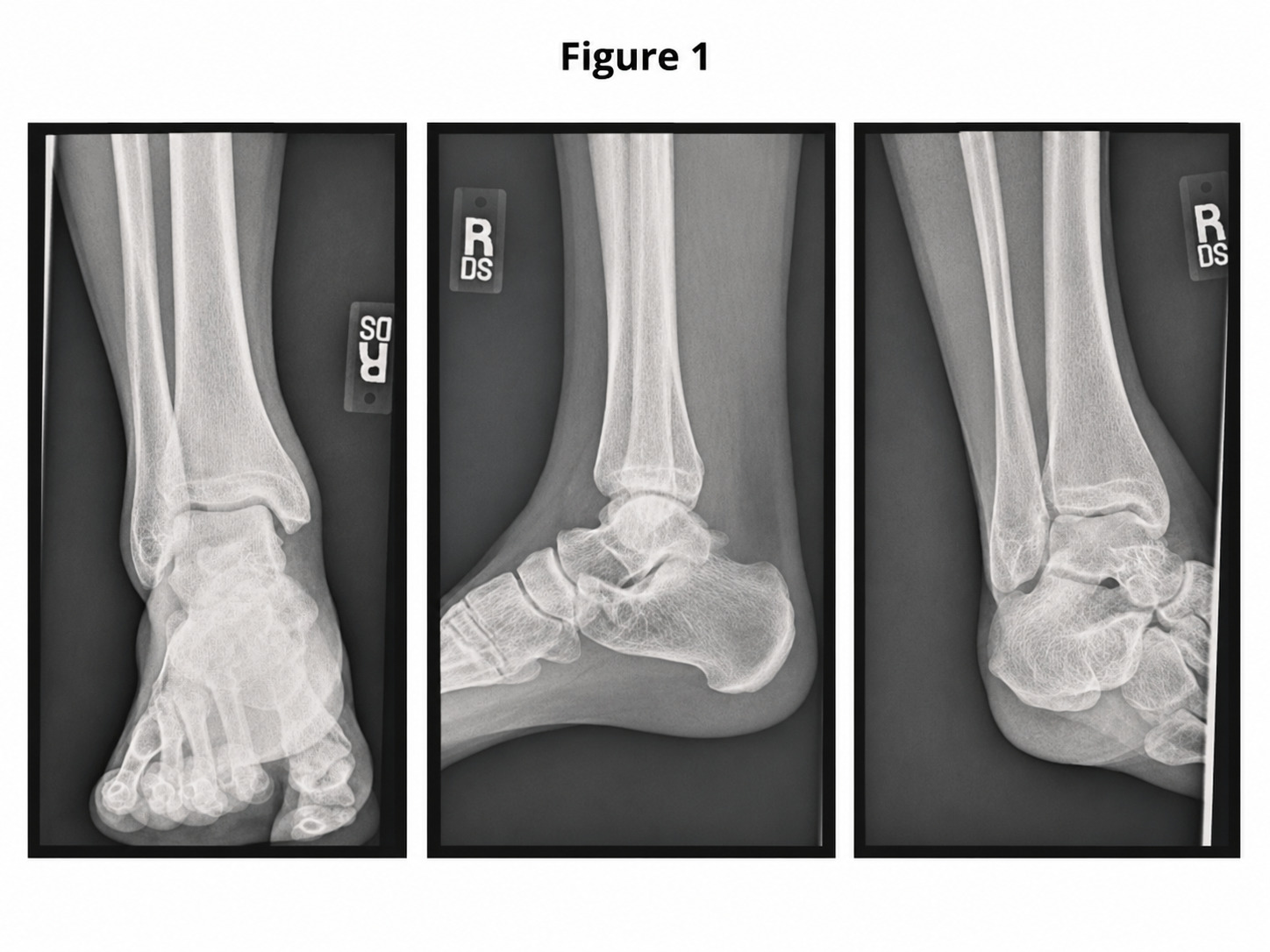
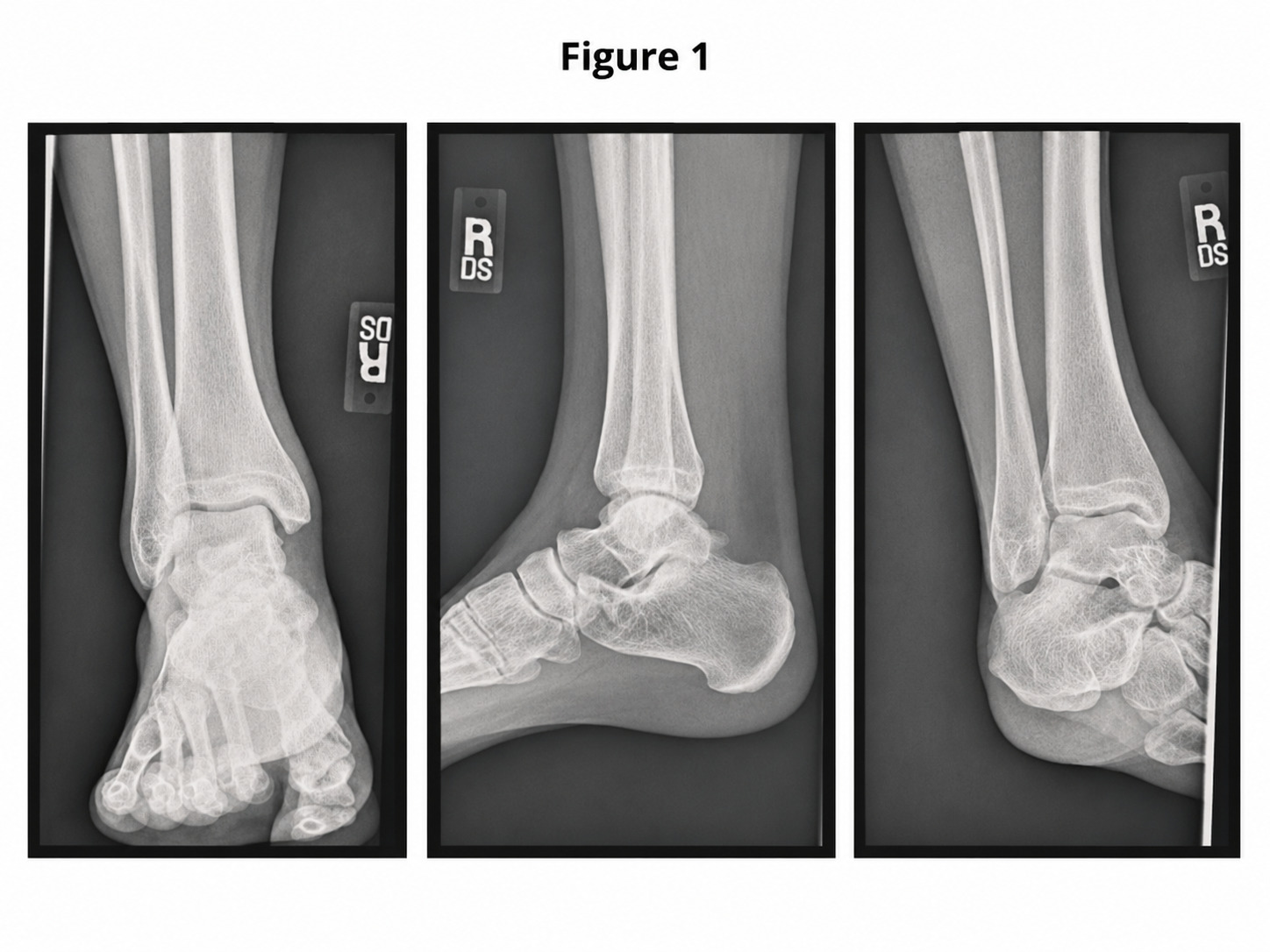
Bilateral ankle radiographs obtained during the first visit of the current episode demonstrated no evidence of acute fracture or osseous pathology (Figure 1). The patient began conservative care consisting of activity modification, soft-tissue interventions, ankle mobility exercise, and early strengthening. After 1 to 2 weeks, he reported worsening focal right tibial pain and reduced running tolerance.
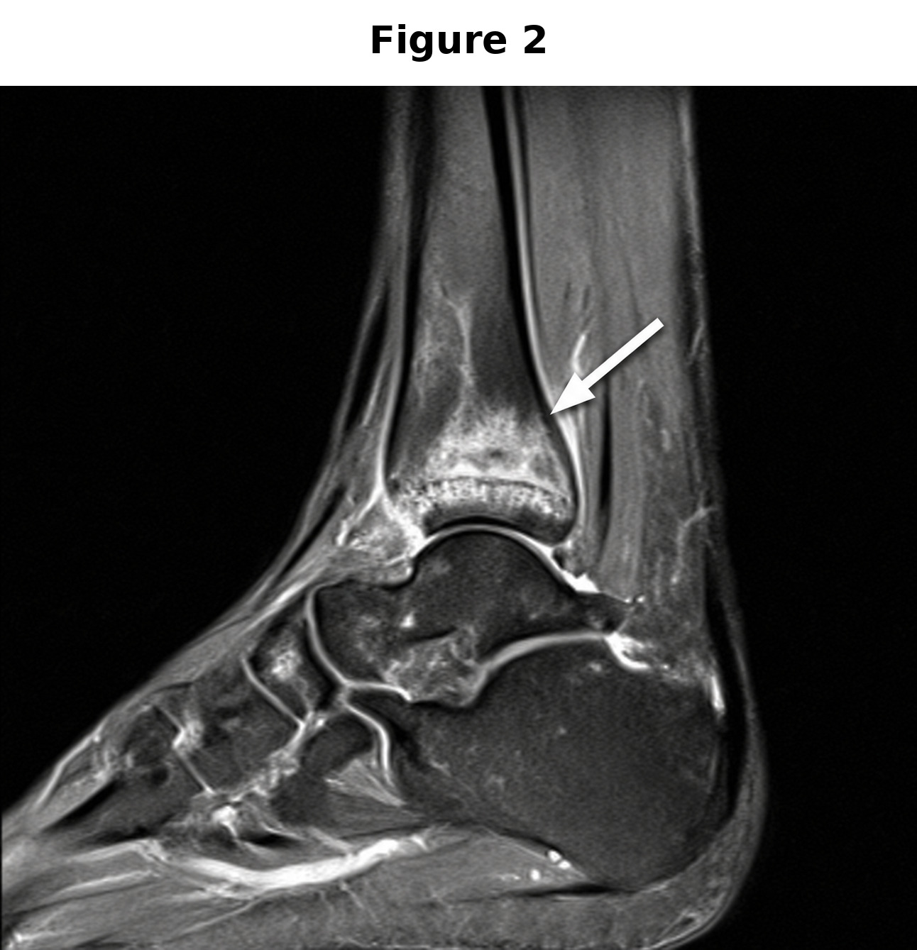
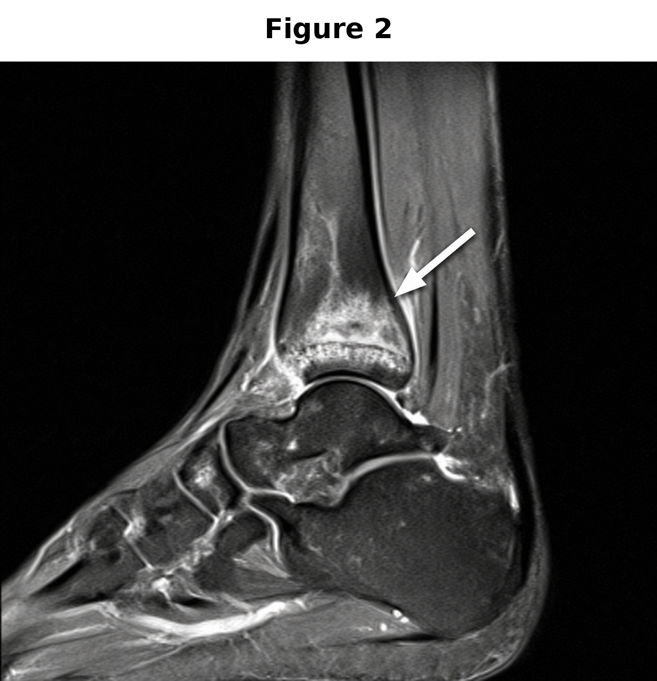
Because of increasing symptom severity and focal distal tibial tenderness despite unremarkable radiographs, magnetic resonance imaging of the right lower leg was ordered. It demonstrated a distal tibial stress fracture with surrounding periostitis and bone marrow edema (Figure 2). Tenosynovitis of the right posterior tibial tendon was also noted, consistent with the prior clinical diagnosis.
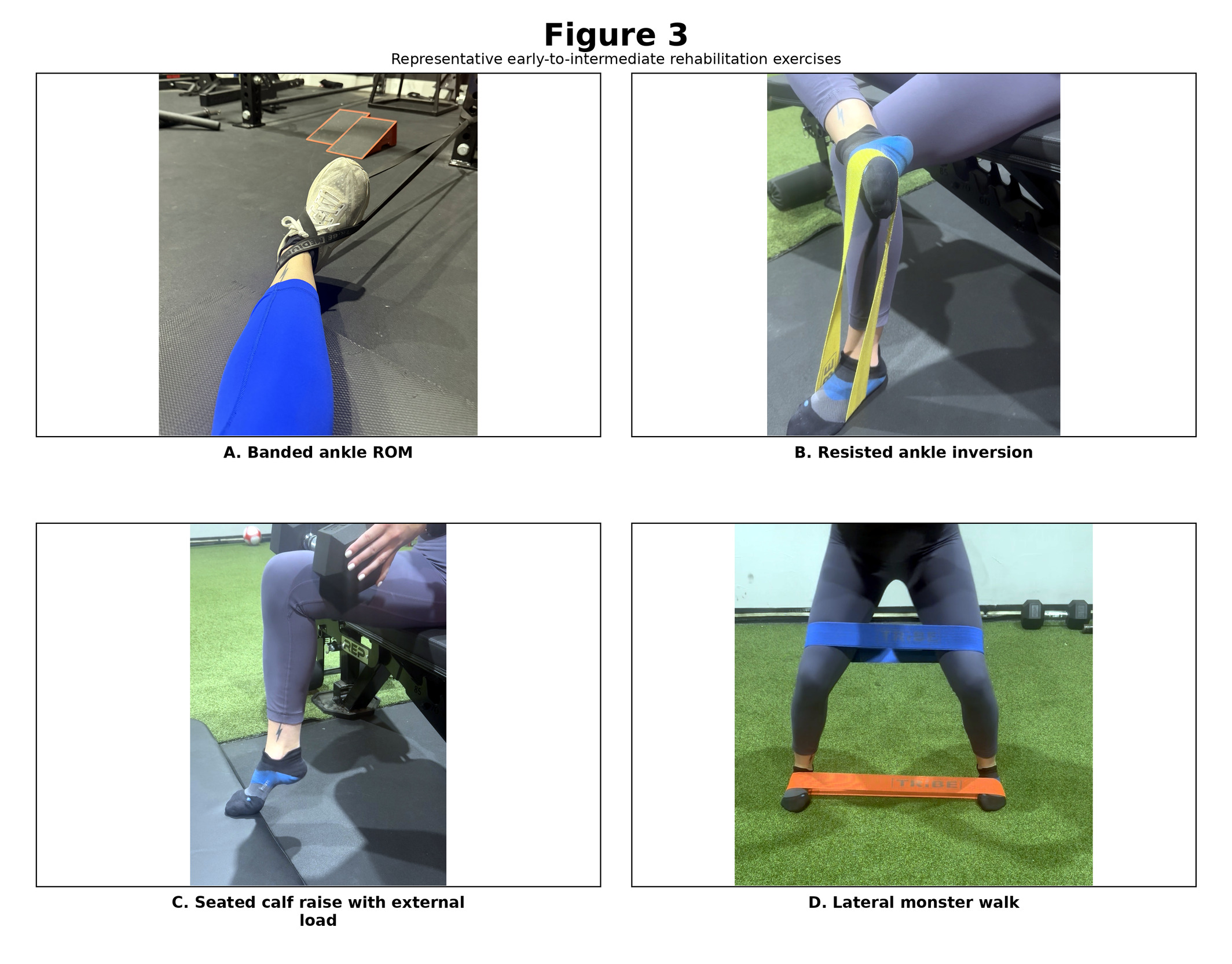
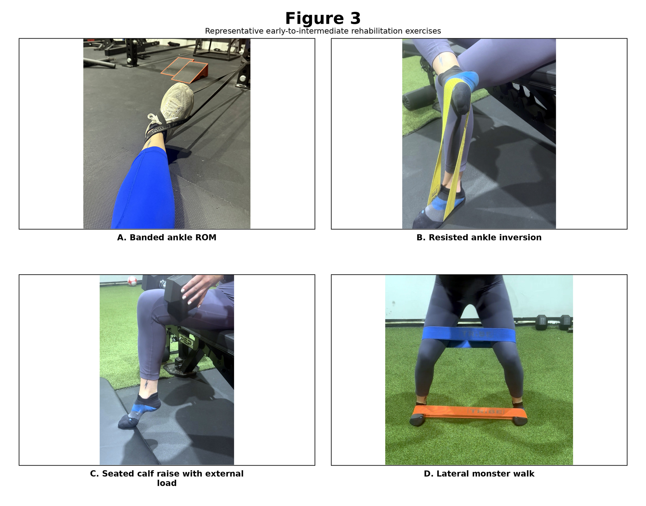
After MRI confirmation, the patient was instructed to discontinue running and reduce weight-bearing volume. As part of orthopedic co-management, he was placed in a walking boot for several weeks to reduce painful loading through the distal tibia. Early rehabilitation included pain-free ankle mobility, resisted ankle inversion, low-load calf and tibialis posterior strengthening, hip abductor and extensor strengthening, adductor activation, and proprioceptive training (Figure 3). Exercises were generally prescribed for 15 repetitions with 5-second isometric holds, while selected calf and adductor exercises were performed for 2 sets of 10 repetitions.
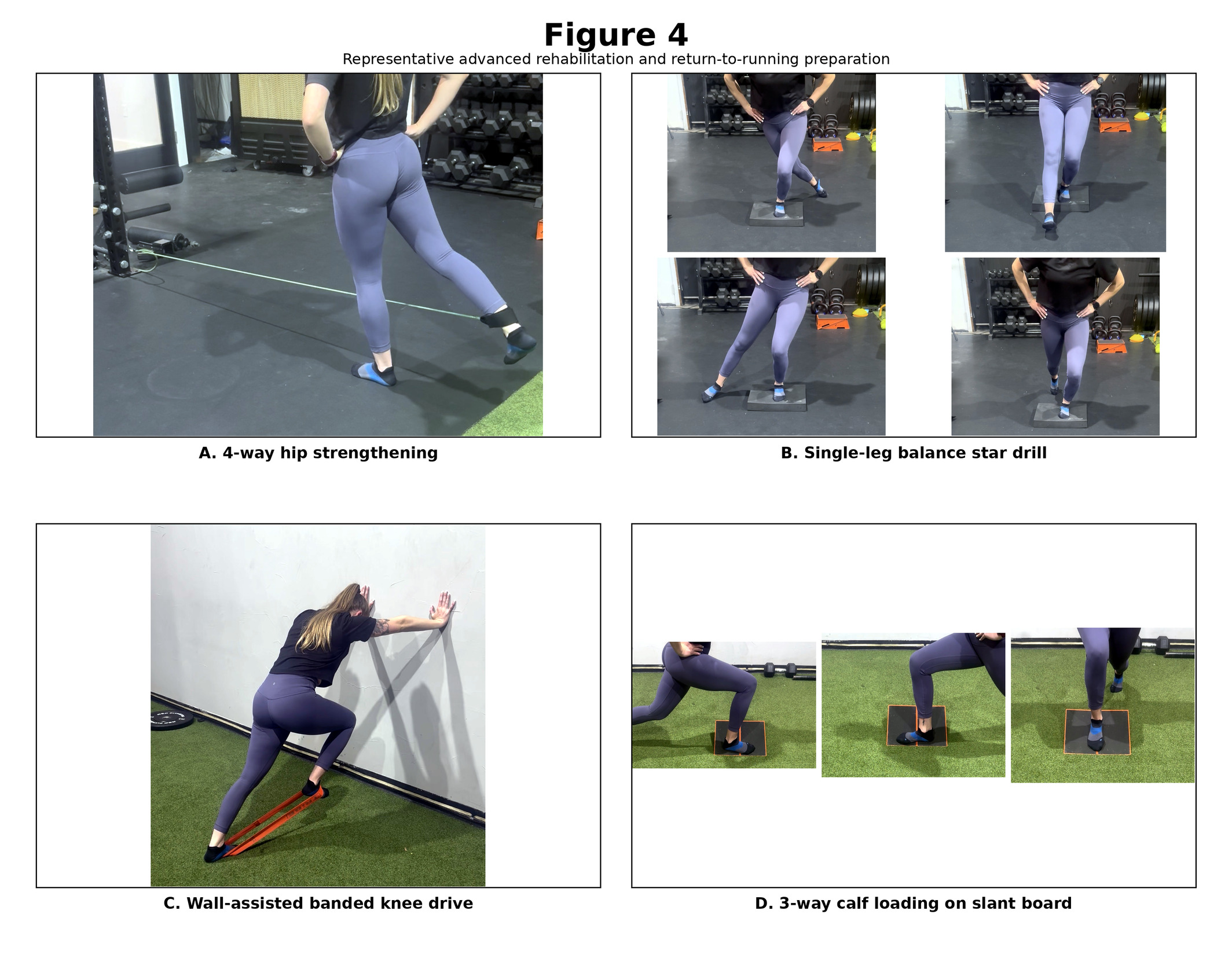
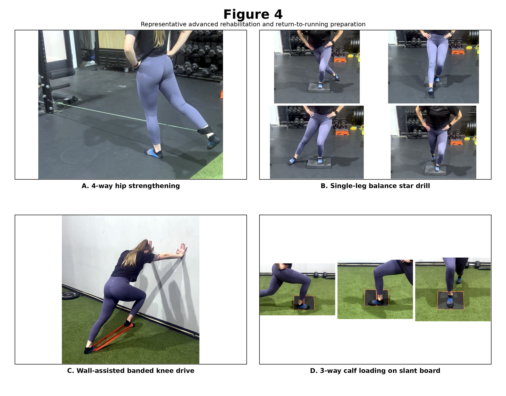
The patient demonstrated gradual reduction in pain during his activities of daily living, with decreased palpatory tenderness along the posterior tibial tendon and distal tibia. Rehabilitation was progressively advanced from supported mobility and activation exercises to dynamic stabilization, eccentric lower-extremity loading, single-leg balance, and multiplanar strengthening. Progressions included ankle circle-board drills, 4-way hip strengthening, step-ups with controlled eccentric step-downs, ball-squeeze bridges, bent-knee calf raises, single-leg balance star drills, lateral band walks, 3-way calf stretching, multiplanar isometric calf raises, banded knee drives, quadruped hip-control exercises, and 90-90 hip mobility drills (Figure 4). The patient completed 24 visits over 3 months (Table 1).
The patient subsequently initiated a self-guided return-to-running program. He reported increasing weekly mileage by approximately 10% for 3 consecutive weeks, followed by a recovery week with approximately 25% lower mileage, and repeating this cycle as tolerated. This progression was patient directed and was not prescribed as a validated protocol. He later returned to the clinic informally and reported completing a 50-kilometer race without recurrence of symptoms. He also reported that a follow-up MRI was unremarkable. The follow-up MRI report and images were not available to the authors for independent review. A formal patient perspective statement was not obtained.
Discussion
Literature Search
A focused narrative literature search was conducted in July 2026 to identify publications addressing tibial bone stress injury, tibial stress fracture, posterior tibial tenosynovitis or tendinopathy, running-related overuse injury, diagnostic imaging, conservative rehabilitation, and return to running. PubMed and Google Scholar were searched using combinations of the following terms: tibial stress fracture, tibial bone stress injury, posterior tibial tenosynovitis, posterior tibial tendinopathy, tibialis posterior, running injury, ultramarathon, distance runner, collegiate distance runner, magnetic resonance imaging, rehabilitation, return to running, acute:chronic workload ratio, and training load. English-language peer-reviewed articles were prioritized, with emphasis on systematic reviews, narrative reviews, cohort studies, consensus or clinical guidance, and publications directly relevant to runners or physically active populations. Reference lists of selected articles were reviewed to identify additional pertinent sources. Because the purpose of the search was to contextualize a case report rather than conduct a systematic review, formal risk-of-bias assessment and quantitative synthesis were not performed.
Review of the Literature
Running-related lower extremity injuries are multifactorial and may be influenced by previous injury, training exposure, tissue capacity, biomechanics, and recovery.1 Bone stress injury represents a continuum of skeletal overload in which repetitive mechanical loading exceeds the capacity for bone repair, producing localized pain and, in more advanced cases, stress fracture.2,3 In runners, the tibia is consistently identified as one of the most common stress-fracture locations, with several reviews and clinical series reporting that tibial injuries account for approximately one-half of running-related stress fractures.3–5
Tibial bone stress injuries are not limited to adult recreational runners. Yagi et al6 prospectively studied high-school runners and reported that medial tibial stress syndrome and tibial stress fracture were common lower-leg disorders in this younger running population. Contemporary collegiate running data also support the clinical relevance of bone stress injury in young high-volume runners. Wolff et al7 reported that bone stress injuries were common in NCAA Division I distance runners, particularly among female athletes, and varied across competitive phases of the season. Sex-related differences have also been described. In a systematic review and meta-analysis of lower-extremity stress-fracture risk factors in runners, Wright et al8 reported that female sex and previous stress fracture history were the only risk factors strongly supported by pooled data. Our case involved a male runner who denied previous fracture, emphasizing that clinicians should not rely solely on demographic risk profiles when evaluating progressive focal tibial pain in endurance athletes.
Training-load change is another important consideration. In runners training for the New York City Marathon, Toresdahl et al9 found that a greater number of days with an acute:chronic workload ratio of 1.5 or greater was associated with increased injury odds, with each additional day above this threshold associated with a 6% increase in injury odds. More recent cohort data suggest that abrupt single-session distance spikes may be particularly relevant. Frandsen et al10 determined that running-related overuse injury risk increased when the distance of a single running session exceeded 10% of the runner’s longest run during the preceding 30 days, whereas week-to-week ratio measures were not associated with injury risk. Together, these studies support a more nuanced interpretation of running progression that considers recent workload history, individual session distance, recovery, symptoms, and functional tolerance rather than weekly mileage alone.9–11
Clinical recognition of tibial bone stress injury can be difficult early in the injury continuum. Symptoms may initially occur only with prolonged running but may later appear earlier during activity, persist after activity, or occur with walking and daily weight-bearing. Focal tibial tenderness, declining running tolerance, and symptoms that persist despite activity modification should increase suspicion for bone stress injury. Plain radiographs are often unremarkable early in the course of stress injury, whereas magnetic resonance imaging can demonstrate periosteal edema, bone marrow edema, cortical involvement, and associated soft-tissue pathology.3,12
The tibialis posterior contributes to inversion and plantar flexion, assists with control of pronation, and supports the medial longitudinal arch during weight-bearing.13 Posterior tibial tendon pathology may produce medial ankle pain, tenderness along the tendon course, and reduced tolerance to repetitive weight-bearing activity. Most literature concerning posterior tibial tendon dysfunction involves middle-aged adults with progressive arch collapse, whereas less has been written about posterior tibial tenosynovitis or tendinopathy in younger endurance athletes without fixed deformity. Available evidence does not establish that posterior tibial tenosynovitis directly causes tibial stress fracture; however, these conditions may coexist in runners exposed to repetitive loading, reduced recovery, muscular fatigue, and altered lower-extremity loading patterns.
Conservative management of posterior tibial tendon symptoms commonly includes activity modification, footwear or orthotic support when indicated, and progressive resistance exercise.5,14,15 Rehabilitation following tibial stress fracture similarly begins with reduction or temporary cessation of painful impact activity and progresses toward restoration of ankle mobility, calf and foot strength, proximal lower-extremity control, balance, and sport-specific loading.5,16 Return-to-running recommendations after tibial bone stress injury are generally criteria-based rather than dependent on a fixed period of rest or a universal mileage rule. Common considerations include pain-free walking, resolution or substantial reduction of focal bony tenderness, restoration of lower-extremity strength, tolerance of functional loading, and management of relevant contributing factors.17
Comparison With the Present Case
This case shares several features with the published literature on tibial bone stress injury in runners, including gradual onset, high repetitive running exposure, increasingly focal tibial tenderness, reduced running tolerance, and unremarkable initial radiographs followed by MRI confirmation. It also differs from several common risk profiles because the patient was male, denied previous fracture, and presented with concurrent posterior tibial tenosynovitis. The overlap between tendon-related medial ankle pain and evolving distal tibial bone stress injury likely contributed to diagnostic complexity.
The transition from diffuse or tendon-course medial ankle pain to deeper and more focal distal tibial pain was the most clinically important change in this case. This change in symptom behavior prompted reconsideration of the working diagnosis and referral for advanced imaging. The case therefore reinforces that persistent focal bony tenderness in a runner should prompt reassessment, particularly when symptoms worsen despite appropriate initial care or occur with previously tolerable running distances.
The rehabilitation program was organized as a graded progression rather than as a collection of isolated corrective exercises. Early care emphasized reduction of impact loading, pain-free ankle mobility, tibialis posterior activation, low-load calf strengthening, and proximal hip activation. This was followed by eccentric step-downs, bent-knee calf raises, resisted hip strengthening, single-leg balance, lateral band walking, multiplanar calf loading, and running-specific control drills. This sequence was intended to restore ankle dorsiflexion, calf and tibialis posterior capacity, hip control, proprioception, and tolerance to progressive loading. The program is consistent with conservative rehabilitation recommendations for posterior tibial tendon symptoms and tibial stress fracture management, although the available evidence does not establish the superiority of any single exercise protocol.5,14–16
The patient later used a self-directed return-to-running strategy that included approximately 10% weekly mileage increases for 3 weeks followed by a recovery week. This approach is reported descriptively and should not be interpreted as a validated or universally protective protocol. Current return-to-running literature after tibial bone stress injury emphasizes individualized progression based on symptoms, injury severity, pain-free walking, focal tenderness, functional loading tolerance, and strength.17 Running-load research further suggests that clinicians should monitor abrupt workload spikes, including high acute:chronic workload days and sudden increases in single-session distance, rather than relying on a single weekly percentage rule.9,10
Limitations
This single case report cannot establish causation or determine the effectiveness of any specific intervention. The rehabilitation program contained multiple concurrent components, preventing attribution of improvement to 1 exercise or treatment. The return-to-running program was self-directed, and adherence and weekly mileage were not independently verified. Although the patient reported that follow-up MRI findings were unremarkable, the report and images were not available to the authors for independent review. He had previously withdrawn from care, which limited continuity of clinical observation. Nutritional status, energy availability, training periodization, and other potential contributors to bone stress injury were based on patient report and were not comprehensively assessed. A formal patient perspective statement was not obtained.
Conclusion
This case report describes a young ultramarathon runner with posterior tibial tenosynovitis and subsequent MRI-confirmed distal tibial stress fracture. The case highlights the need to reconsider the working diagnosis when high-volume runners develop worsening focal tibial pain, declining running tolerance, or an unexpected response to conservative care. Normal radiographs do not exclude early bone stress injury, and MRI may be warranted when suspicion remains high. A graded rehabilitation program addressing ankle mobility, calf and tibialis posterior capacity, proximal lower-extremity control, balance, and progressive impact exposure was followed by a self-directed return to running. The patient later reported completing a 50-kilometer race and an unremarkable follow-up MRI; however, these outcomes were self-reported and the imaging was not independently reviewed.
Artificial Intelligence Disclosure
Generative artificial intelligence was used to assist with language editing, organization, and submission formatting. It was not used as an author or as a primary source. The authors reviewed, verified, and take responsibility for all content, citations, interpretations, and conclusions.
Conflicts of Interest
The authors declare no financial or nonfinancial conflicts of interest related to this work.
Funding
No external funding was received for this work.
Consent for Publication
Written informed consent was obtained from the patient for publication of this deidentified case report and associated clinical images.
Ethics Statement
This report describes information obtained during routine clinical care and does not report a prospective research intervention. Written informed consent was obtained from the patient for publication of deidentified clinical information and associated images. Institutional review board review was not required for this single-patient case report according to institutional policy. The rehabilitation photographs are representative demonstrations and do not depict the patient.