Loading [Contrib]/a11y/accessibility-menu.js
1.
Lopes M, Coleman R. IMAGING RECOMMENDATIONS IN GUIDELINES RELEVANT TO CHIROPRACTIC CARE: A SYSTEMATIC REVIEW OF THE AVAILABILITY OF DIRECT COMPARATIVE EVIDENCE. JCC. 2026;9(1):382-396.
Download all (3)

Abstract

Objective

To systematically review clinical guidelines and related literature to determine whether the evidence supporting recommendations affecting the use of imaging in chiropractic practice includes studies that have directly compared outcomes of chiropractic care guided by radiographic findings versus chiropractic care delivered without radiographic guidance.

Methods

A systematic search of PubMed, Google Scholar, Index to Chiropractic Literature, Cochrane Library, and Guideline repositories was performed for English language publications between January 2007 and March 2026. Searches used combinations of keywords related to chiropractic practice, spinal imaging, radiography, spinal pain, and imaging recommendations. Chiropractic imaging guidelines, consensus statements, systematic reviews, rapid reviews, and narrative reviews addressing spinal imaging relevant to chiropractic practice were eligible for inclusion. Included publications and their reference lists were examined to identify studies directly comparing outcomes of chiropractic care in which radiographic findings informed assessment or treatment planning versus chiropractic care delivered without radiography informed management. Studies that did not address spinal imaging recommendations were excluded.

Results

A total of 249 publications met initial screening criteria and 26 guidelines and review articles were included after full text evaluation. These guidelines commonly recommended selective imaging based primarily on the presence of red flags, suspicion of pathology, or anticipated changes in patient management. Across the guidelines included and their cited reference lists, no direct comparative clinical trials were identified evaluating chiropractic care delivered with radiography guided biomechanical analysis versus comparable care delivered without radiographic guidance. The evidence cited in support of guideline recommendations primarily consisted of studies addressing diagnostic yield, prevalence of radiographic findings, radiation exposure risk modeling, and healthcare utilization patterns rather than comparative clinical outcome trials.

Conclusion

Within the guideline and review literature examined, direct comparative evidence evaluating the clinical effectiveness of radiography guided chiropractic care versus care delivered without such imaging guidance was not identified. Accordingly, the existing guideline literature neither confirms nor refutes the effectiveness of radiography guided biomechanical decision making in chiropractic practice. This represents an evidence gap rather than evidence of ineffectiveness. Future controlled comparative studies are needed to determine whether imaging informed biomechanical assessment influences patient outcomes in chiropractic care.

Introduction

Clinical practice guidelines are intended to synthesize available research evidence to support clinical decision making and promote consistent standards of care. Within chiropractic practice, spinal radiography has historically been used for multiple purposes, including detection of pathology, assessment of structural abnormalities, identification of potential contraindications to spinal manipulation, and biomechanical or postural assessment of the spine.1–4

Over the past 2 decades, several national and international guideline efforts have addressed the role of spinal imaging in the management of spinal pain conditions within chiropractic and medical healthcare settings. These guidelines generally recommend against initial imaging prior to chiropractic care and instead support selective imaging when serious underlying pathology or other clinical red flags are suspected, including significant trauma, suspected malignancy, infection, and severe or progressive neurological deficits.5–10

However, clinical guidelines are inherently selective in the evidence they incorporate. Evidence based guideline development processes typically place greater weight on higher certainty evidence, particularly systematic reviews and randomized controlled trials.11,12 This process may under-represent other forms of evidence, including observational research, mechanistic reasoning, clinical expertise, and patient specific factors. While this prioritization is intended to reduce bias, it may also result in a narrower representation of the broader body of scientific and clinical knowledge relevant to clinical decision-making.13,14

An additional limitation of guideline development arises when direct comparative outcome studies addressing a specific clinical question are absent. In such situations, recommendations may rely on indirect evidence, judgments regarding potential benefits and harms, or consensus interpretation of limited data.11,15 The absence of direct evidence can therefore create uncertainty regarding the validity of recommendations that either support or discourage particular clinical practices.

Within chiropractic imaging debates, an important unanswered question is whether the use of radiography to inform biomechanical assessment and treatment planning influences clinical outcomes compared with chiropractic care delivered without such imaging guidance. Some chiropractic practitioners use radiographic assessment to guide spinal manipulative therapy (SMT) and other biomechanically oriented treatment approaches,2,3 whereas many contemporary guidelines recommend against routine radiographic imaging for patients with non-specific spinal pain in the absence of “red flags” or other concerning clinical indications.5–10

To date, it remains unclear whether guideline recommendations regarding spinal imaging in chiropractic are supported by direct comparative evidence evaluating clinical outcomes of care delivered with versus without radiography guided decision-making.

The purpose of this study was therefore to systematically review chiropractic imaging guidelines and related literature to determine the evidentiary basis supporting their recommendations. Specifically, this review sought to identify whether direct comparative studies exist that evaluate clinical outcomes of chiropractic delivered with radiography guided biomechanical assessment compared with similar care delivered without radiographic guidance.

Methods

Study Design

This systematic review was conducted according to general methodological principles described in the Preferred Reporting Items for Systematic Reviews and Meta-Analyses (PRISMA) recommendations.16 The objective was to evaluate guideline and review literature addressing spinal imaging recommendations relevant to chiropractic practice and to determine whether evidence existed directly comparing chiropractic care delivered with radiography guided treatment planning versus chiropractic care delivered without imaging guidance.

Eligibility Criteria

Publications were eligible for inclusion if they met all the following criteria:

  1. Published in English.

  2. Addressed spinal imaging in chiropractic practice or spinal pain management relevant to chiropractic care.

  3. Included discussion of imaging recommendations, imaging appropriateness, diagnostic imaging utilization, or imaging related clinical decision making.

  4. Were clinical practice guidelines, consensus statements, systematic reviews, rapid reviews, narrative reviews with literature synthesis, imaging appropriateness guidelines, or related evidence summaries.

  5. Were published between January 2007 and March 2026.

The starting year of 2007 was selected because the publication of the Bussières imaging guidelines in 20081 represented 1 of the earliest major evidence-based chiropractic imaging guideline initiatives and substantially influenced subsequent literature and guideline development.

Publications were excluded if they:

  1. Focused exclusively on imaging modalities unrelated to spinal radiography without discussion of radiographic decision making;

  2. Did not address spinal imaging recommendations;

  3. Consisted solely of editorials, opinion papers, or commentaries lacking literature review components; were case reports or primary diagnostic studies without guideline or evidence synthesis objectives.

Information Sources

Literature searches were performed using publicly accessible databases and guideline repositories. Sources included:

PubMed, Google Scholar, Cochrane Library, Index to Chiropractic Literature and Guideline repositories including National Institute for Health and Care Excellence, American College of Radiology and World Health Organization.

Reference lists of all included publications were manually screened to identify additional relevant studies.

Search Strategy

Searches used combinations of keywords related to chiropractic practice, spinal imaging, radiography, spinal pain, and imaging recommendations.

Representative search concepts included:

Chiropractic, spinal manipulation, manual therapy, spinal imaging, radiography, X-ray, low back pain, neck pain, clinical guideline, practice guideline, systematic review, imaging appropriateness

Search terms were combined using Boolean operators where supported by the database platform.

A representative PubMed search strategy was:
(chiropractic OR “spinal manipulation” OR “manual therapy”) AND (imaging OR radiography OR “x-ray” OR radiograph*) AND (spine OR “low back pain” OR “neck pain”) AND (guideline OR “practice guideline” OR “systematic review” OR review)

Searches were conducted by one reviewer and were restricted to English-language publications and the period January 2007 through March 2026.

Study Selection

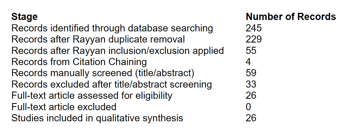
The literature search identified 245 records across all databases, which were imported into Rayyan (Qatar Computing Research Institute, Doha, Qatar), a web-based application designed for semi-automated systematic review screening. Within Rayyan, 16 duplicate records were identified and removed, leaving 229 records for automated title and abstract screening using the predefined inclusion and exclusion criteria. Four additional articles were identified through citation chaining, bringing the total number of records identified during the search process to 249.

Following duplicate removal and initial screening in Rayyan, 55 potentially eligible publications remained. These 55 records, together with 4 additional articles identified through citation chaining, were independently reviewed by both authors at the title and abstract level. Thirty-three of the 59 publications were excluded because they did not meet the eligibility criteria. The remaining 26 publications (Appendix A) underwent full-text review (Figure 1).

Full-text articles were imported into Sharly AI (Vox AI Inc.) and docAnalyzer.ai (AI For Verticals, Inc.) as organizational tools to facilitate review of article content and study characteristics. Each article was independently reviewed in full by both authors, including examination of cited references. Eligibility determinations, study classification, data extraction, and interpretation of findings were performed by the authors and were not delegated to artificial intelligence software.

Data Extraction

Data extracted from each included publication included:

Author and year; publication type; clinical scope; imaging focus; role within the evidence base; imaging recommendations; evidence type; presence or absence of direct comparative evidence.

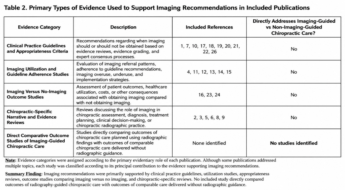
Evidence categories identified during synthesis included:

  1. Clinical practice guidelines and appropriateness criteria.

  2. Imaging utilization and guideline adherence.

  3. Imaging versus no imaging outcome studies.

  4. Chiropractic specific narrative and evidence reviews.

  5. Direct comparative outcome studies of imaging guided chiropractic care.

Comparative Evidence Assessment

Included articles were reviewed to determine whether direct comparative clinical outcome evidence existed, searching for studies directly comparing chiropractic care delivered with radiography guided treatment planning versus chiropractic care delivered without radiography guidance.

Evidence Synthesis

Because the objective of the review was to characterize guideline recommendations and the evidence supporting those recommendations rather than pool homogeneous treatment effects, findings were synthesized narratively rather than by quantitative meta-analysis.

Special emphasis was placed on distinguishing:

Direct comparative outcome evidence, diagnostic evidence, utilization studies, imaging appropriateness literature, and chiropractic specific biomechanical or technique related publications.

Artificial intelligence software (Chat GPT/Open AI) was used to assist with table formatting and suggesting methods for evidence classification. All study selection, data extraction and evidence classification were performed by the authors. Evidence classification suggestions were reviewed, verified and finalized by the authors using the original publications. The AI system was not used to determine study eligibility, perform critical appraisal, assign final evidence classifications without author review, or draw study conclusions.

Results

The evidence supporting imaging recommendations primarily consisted of clinical practice guidelines, appropriateness criteria, utilization and cost studies, outcome studies comparing imaging versus no imaging, association studies, radiation exposure modeling and biomechanical reliability investigations. No included publication directly compared outcomes of chiropractic care delivered using radiography guided treatment planning with outcomes of comparable chiropractic care delivered without radiographic guidance.

No randomized trials or controlled comparative investigations directly evaluating radiography guided chiropractic management versus non-imaging guided chiropractic management were identified among the included literature set. Table 1 shows the characteristics of included publications. Table 2 shows the types of evidence used to support imaging recommendations.
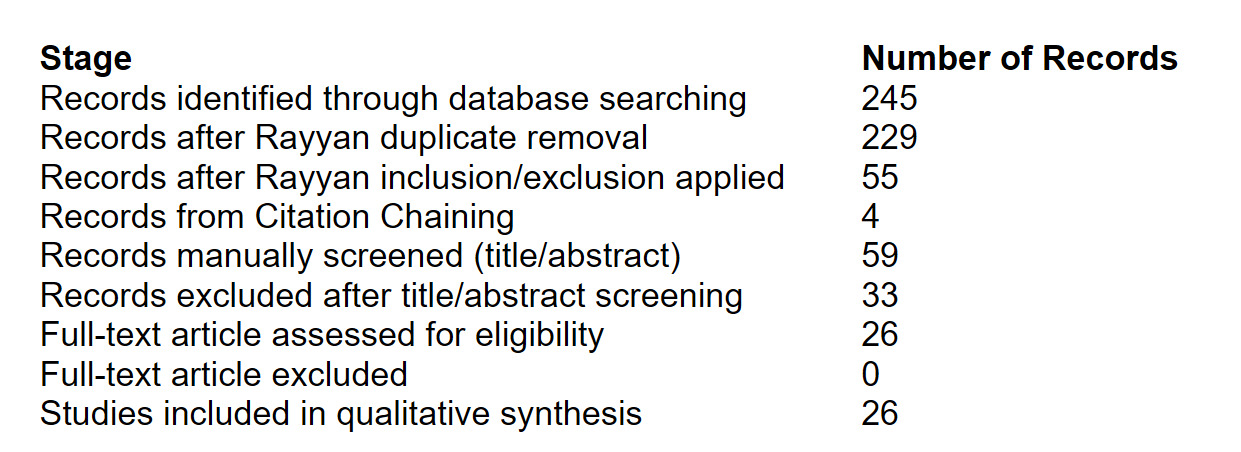
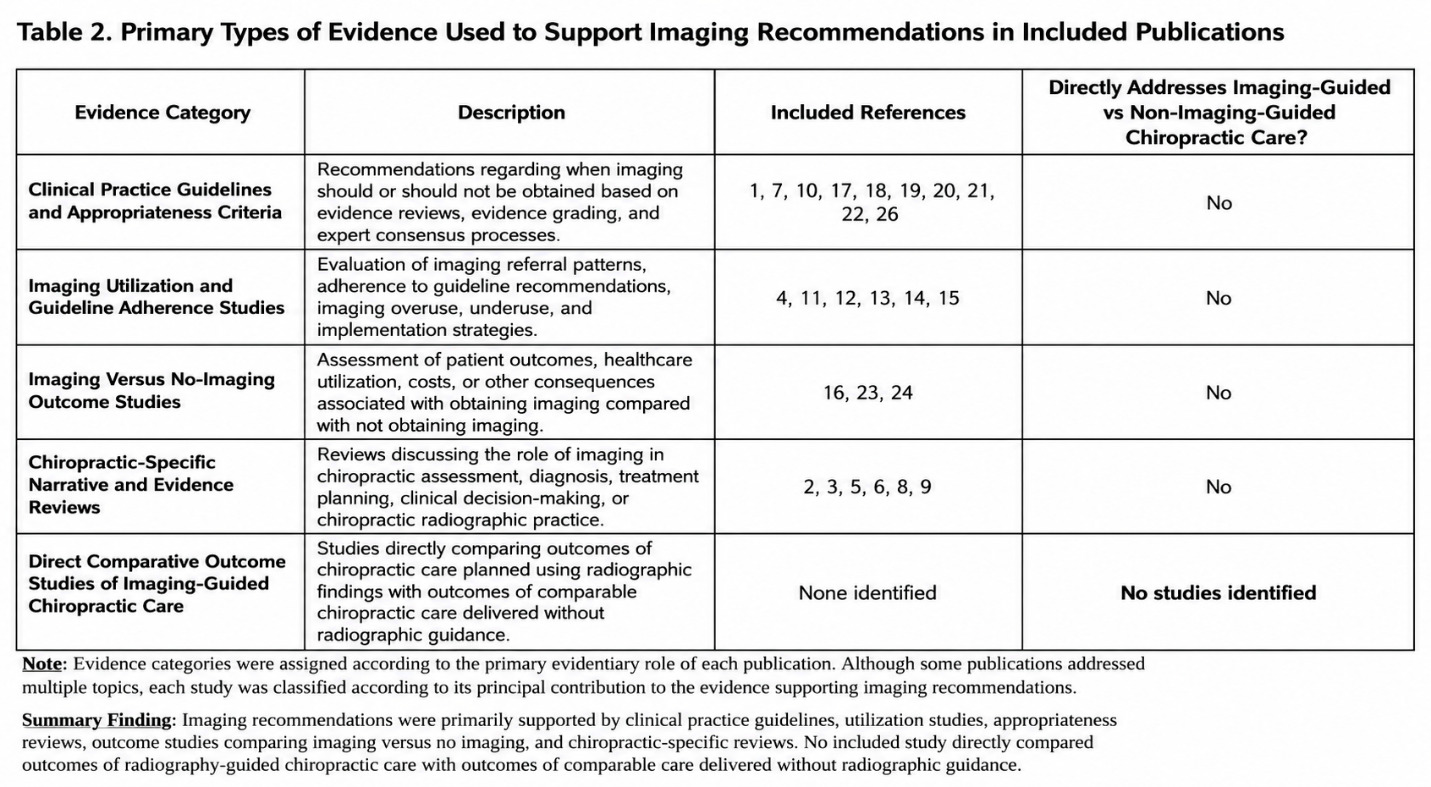
Figure 1
Figure 1.Flow Diagram Data
Table 1.Characteristics of Included Publications and Assessment of Direct Comparative Evidence
Ref Author/Year Type Scope Imaging Focus Addresses Direct Imaging-Guided vs Non-Imaging-Guided Chiropractic Outcomes?
1 Stochkendahl 2018 Guideline LBP/Radiculopathy Imaging indications No
2 Jenkins 2018 Narrative Review Chiropractic Spinal X-ray use No
3 Williams 2024 Practice Study Chiropractic Routine radiography No
4 Jenkins 2018 Systematic Review Low back pain Guideline concordance No
5 Triano 2013 Review SMT site selection Treatment planning No
6 Corso 2020 Rapid Review Chiropractic Routine radiographs No
7 Bussières 2008 Guideline Spinal disorders Radiography No
8 Taylor 2012 Narrative Review Elderly patients Spinal imaging No
9 Hawk 2017 Consensus/SR Older adults Best practices No
10 Whalen 2022 Guideline Mechanical LBP Management/imaging No
11 Wáng 2018 Narrative Review Low back pain Appropriate imaging No
12 Flynn 2011 Review Low back pain Imaging harms No
13 Belavy 2022 SR/MA Low back pain Reduce low-value imaging No
14 Logan 2019 SR/MA Primary care Appropriateness No
15 Dagenais 2014 Systematic Review LBP care Imaging utilization No
16 Lemmers 2019 Systematic Review Low back pain Imaging vs no imaging No
17 Monticone 2013 Guideline Neck pain Imaging indications No
18 Globe 2016 Guideline Chiropractic LBP Care including imaging No
19 Chou 2007 Guideline Low back pain Diagnostic imaging No
20 Chou 2011 High-Value Care Low back pain Diagnostic imaging No
21 Hutchins 2021 ACR Guideline Low back pain Appropriateness No
22 Côté 2016 Guideline Neck pain Imaging indications No
23 Chou 2009 SR/MA Low back pain Imaging strategies No
24 Karel 2015 Meta-analysis MSK disorders Routine imaging No
25 Cuff 2020 Scoping Review MSK pain Imaging guidelines No
26 de Campos 2017 Guideline Summary LBP/Sciatica NICE imaging context No

Abbreviations: LBP = low back pain; SR = systematic review; MA = meta-analysis; SMT = spinal manipulative therapy; ACR = American College of Radiology.
Key finding: No included publication directly compared outcomes of chiropractic care guided by radiographic findings with comparable chiropractic care delivered without radiographic guidance. Note: Reference numbers correspond to publications included in the evidence synthesis. Full bibliographic information for all included studies is provided in Appendix A.

Discussion

Major clinical practice guidelines commonly discourage routine imaging in patients with acute low back pain (LBP) in the absence of “red flag” findings.6–8,17 This position is largely based on evidence demonstrating that many imaging abnormalities occur in asymptomatic individuals and concerns that routine imaging of broad primary care non-specific low back pain populations has not consistently been associated with improved average patient outcomes.18–21 However, these recommendations are primarily derived from management paradigms emphasizing passive medical care, reassurance, medication, and time-dependent recovery rather than mechanically targeted interventions involving repeated high-velocity, low-amplitude (HVLA) force application. However, some studies in chiropractic patient populations have also failed to demonstrate improved clinical outcomes associated with routine initial imaging.22,23

Nevertheless, experimental evidence suggests that variations in thrust force and thrust duration may influence vertebral displacement, muscular response amplitude, and muscle spindle activity, although the biomechanical details of SMT/HVLA remain incompletely understood.24,25 This distinction is clinically important. Chiropractic HVLA procedures involve directed mechanical loading of spinal tissues and assume a degree of structural integrity, biomechanical predictability, and force tolerance. In this context, radiographic imaging may plausibly serve purposes extending beyond identification of a singular nociceptive source. Plain radiography may contribute contextual information regarding spinal alignment, transitional anatomy, degenerative change, spondylolisthesis, scoliosis, instability suspicion, congenital variation, or other structural features that may inform clinical assessment, management decisions, contraindication assessment, risk stratification, or, in some chiropractic approaches, procedural planning.26–31

A related consideration is that “non-specific” low back pain accounts for approximately 90% of low back pain presentations and, by definition, cannot be confidently attributed to a specific identifiable pathology or nociceptive source.32 Like low back pain, most patients presenting with neck pain have non-specific neck pain, and the underlying etiological factors are often poorly understood and multifactorial.33 Further, the observation that many imaging findings occur in asymptomatic individuals is well supported; however, the clinical significance of such findings depends on their interpretation within the broader context of patient presentation, age, history, examination findings, and clinical decision-making.34 Degenerative findings, disc-space narrowing, facet arthrosis, or alignment variations may lack specificity as isolated pain generators, yet this does not necessarily render them clinically irrelevant.26,28 Imaging findings may still possess biomechanical, structural, or prognostic significance even when they are not independently diagnostic of pain causation.30,31 Furthermore, asymptomatic prevalence studies do not by themselves establish that structural findings are unrelated to future disability, recurrent pain episodes, altered load tolerance, or treatment responsiveness in all cases. Some imaging findings may demonstrate prognostic associations with future pain or disability outcomes, although these relationships are often found in studies to be modest and heterogeneous.18,35

Prognostic Considerations

The rationale common in guidelines for delaying imaging for approximately 6 weeks for non-red flags cases also warrants contextual consideration. This recommendation is generally based on the observation that many patients improve symptomatically during the acute phase and that routine imaging does not improve average short-term outcomes in nonspecific LBP populations.6–8,17 However, contemporary prognosis literature demonstrates that acute LBP is not uniformly benign or reliably self-limited. Substantial proportions of patients develop persistent, recurrent, or disabling symptoms despite early symptomatic improvement.36–41 Systematic reviews and longitudinal cohort studies have shown that recurrence rates are common and that early improvement does not reliably predict long-term resolution.36–38,40

Accordingly, symptom improvement alone may be an insufficient endpoint when repeated mechanical interventions are planned. If structural abnormalities relevant to force application, joint loading, or spinal stability remain unidentified, it may be reasonable to consider whether imaging could be helpful in selected patients undergoing prolonged empiric HVLA treatment. This does not imply that all patients with acute LBP require radiography, nor does it establish that imaging guided HVLA treatment is superior in randomized trials. Rather, it highlights an important evidence gap: studies informing recommendations against routine imaging did not specifically assess whether radiographic information improves outcomes when clinicians deliver mechanically targeted manual therapies. The literature reviewed in our current study did not identify direct comparative clinical trials evaluating outcomes of mechanically oriented chiropractic care delivered with versus without radiographic guidance.

The absence of direct evidence supporting universal imaging should therefore not be conflated with evidence demonstrating universal lack of utility. Current evidence generally supports selective, clinically reasoned imaging strategies rather than routine imaging.8,42,43 Nevertheless, given the relatively low radiation exposure associated with plain radiography compared with advanced imaging modalities,44 selective radiographic evaluation may be considered as part of individualized clinical decision making when the expected biomechanical, prognostic, or safety related value may outweigh the very small and uncertain theoretical radiation risk associated with low-dose diagnostic imaging.8,45–47

Clinical Reasoning

Under these conditions of uncertainty, and until stronger direct comparative evidence becomes available, a Bayesian-style approach may offer one way to integrate existing evidence, clinical expertise, patient-specific factors, and evolving information in individualized clinical decision making.48 Statements in many chiropractic guidelines and reviews that no evidence was found for imaging beyond red flag indications49 may warrant careful interpretation, because expert opinion is recognized within formal evidence hierarchies as a low-level form of evidence rather than a complete absence of evidence.50 Expert opinion may help inform clinical reasoning in areas where empirical investigations have not yet provided definitive results.50 Such an approach emphasizes clinical context and probability-based reasoning in situations where direct comparative evidence remains limited. A Bayesian-style approach may therefore be one reasonable way to integrate multiple sources of information when direct comparative evidence remains limited.51,52

Guideline Adherence

This ongoing evidence gap may be one factor contributing to variation between current guideline recommendations and imaging practices in chiropractic care. Many chiropractic clinicians do not consistently follow current imaging guidelines; for example, one survey of Australian chiropractors found that 68% reported ordering x-rays in scenarios where guidelines did not recommend imaging,53 while a separate best-evidence synthesis reported overall guideline adherence of 73% among chiropractors.54 In this context, a practical framework that integrates current evidence, established guidance, and clinical decision-making considerations may assist clinicians in applying imaging recommendations within individualized patient care.

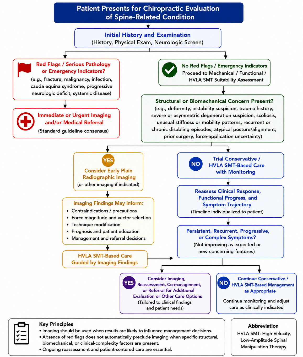
Ultimately, these clinical decisions are made for individual patients rather than for population-level averages upon which much of the literature is based. Accordingly, guideline recommendations derived from frequentist analyses of average group responses may not fully account for the circumstances of every patient at the point of care. With this context in mind, we present the decision-making flow chart in Figure 2 as a practical framework to support individualized consideration of imaging when spinal manipulative therapy using HVLA forces is selected for the management of spinal-related conditions. This framework is proposed as a transparent, hypothesis-generating, expert-informed aid in an area where direct evidence remains limited.
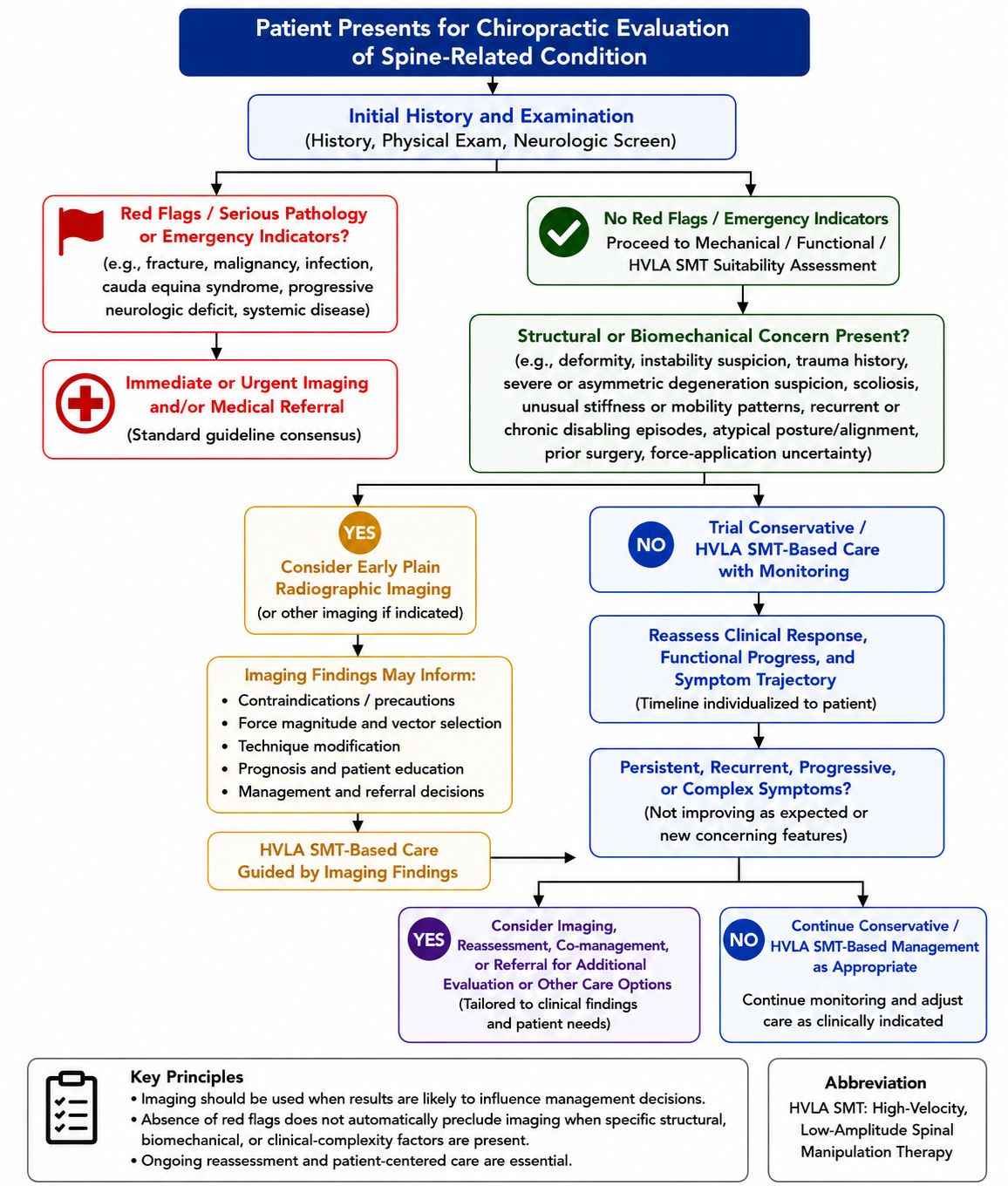
Figure 2
Figure 2.Decision Making Flow Chart

Future Research

The absence of direct comparative studies highlights an important opportunity for future research. Ideally, the question of whether imaging before spinal manipulation improves patient outcomes should be addressed through multiple well-designed studies with adequately large samples. In practice, however, even a single informative study would be challenging to design because the potential clinical value of imaging depends not simply on whether imaging is obtained, but on how imaging findings are interpreted and incorporated into treatment planning. Accordingly, studies designed to evaluate imaging guided care should ensure that imaging findings are used in a manner capable of meaningfully influencing clinical decision making and treatment delivery, rather than functioning solely as a descriptive component of the evaluation process.

From a biomechanical perspective, this issue is complex. The spine is a load-bearing biological structure in which alignment, geometry, material properties, segmental motion, and force transmission influence mechanical behavior. If radiography is hypothesized to improve biomechanical assessment and treatment planning, studies designed to evaluate that hypothesis should consider the specific imaging findings intended to inform clinical decision-making. Depending on the treatment model being evaluated, these may include variables such as regional and intersegmental alignment, transitional anatomy, facet orientation, degenerative findings, or other structural characteristics. These factors may influence biomechanical assessment and treatment planning and therefore should be considered in studies designed to evaluate the clinical utility of imaging guided care. Because spinal manipulation is a mechanical intervention, evaluations of imaging guided care should account for whether radiographic information influences force selection, treatment approach, contraindication assessment, or other clinically relevant decisions.

A further consideration is that the spine is a dynamic biological structure that undergoes adaptation over time. If radiography is hypothesized to improve biomechanical assessment and treatment planning, studies designed to evaluate that hypothesis should incorporate outcome measures that reflect the proposed mechanism of benefit. Although pain intensity is an important patient centered outcome, additional measures may also be relevant, including function, disability, recurrence rates, healthcare utilization, progression of structural findings, and longer-term clinical course. Accordingly, future investigations evaluating the clinical utility of imaging-guided treatment planning should consider outcomes beyond short-term pain reduction alone.

Limitations

This review focused on chiropractic imaging guidelines and related review-level literature rather than on an exhaustive search of all primary studies addressing spinal imaging in musculoskeletal care. Although the reference lists of included guidelines and reviews were examined to identify potentially relevant comparative studies, eligible primary studies may exist outside the sources captured by that approach. The search was limited to English-language publications and to the period from January 2007 through March 2026, which may have excluded earlier or non-English literature. Finally, the finding that direct comparative outcome studies were not identified within the guideline literature should not be interpreted as definitive evidence that such studies do not exist in the broader scientific literature. Rather, the present review indicates that the guidelines and reviews examined did not incorporate direct comparative trials evaluating chiropractic care delivered with versus without radiography guided decision making when formulating their recommendations.

Conclusion

The imaging guideline literature applicable to chiropractic published between January 2007 and March 2026 generally emphasizes imaging for the identification of red flags and other specific clinical indications, while providing comparatively limited consideration of imaging used for biomechanical assessment and treatment planning in patients with non-specific spinal pain. However, the evidence cited in support of these recommendations primarily addresses diagnostic yield, symptom outcomes, potential imaging related harms, and healthcare utilization rather than comparative clinical outcomes associated with biomechanically guided chiropractic care.

Within the guidelines and review literature examined in this study, no direct comparative clinical studies were identified that evaluated chiropractic care delivered with radiography guided biomechanical decision making versus comparable care delivered without radiographic guidance.

Consequently, the current guideline literature neither confirms nor refutes the clinical effectiveness of radiography guided chiropractic care. This represents an important evidence gap that warrants future investigation through well designed comparative studies.


Accepted: August 13, 2026 CDT

References

1.
Bussières AE, Taylor JA, Peterson C. Diagnostic imaging practice guidelines for musculoskeletal complaints in adults-an evidence-based approach-part 3: spinal disorders. J Manipulative Physiol Ther. 2008;31(1):33-88. doi:10.1016/​j.jmpt.2007.11.003
Google Scholar
2.
Jenkins HJ, Downie AS, Moore CS, French SD. Current evidence for spinal X-ray use in the chiropractic profession: a narrative review. Chiropr Man Therap. 2018;26:48. doi:10.1186/​s12998-018-0217-8
Google Scholar
3.
Triano JJ, Budgell B, Bagnulo A, Roffey D, Bergmann TF, Cooperstein R, et al. Review of methods used by chiropractors to determine the site for applying manipulation. Chiropr Man Therap. 2013;21(1):36. doi:10.1186/​2045-709X-21-36
Google Scholar
4.
Taylor JA, Bussières AE. Diagnostic imaging for spinal disorders in the elderly: a narrative review. Chiropr Man Therap. 2012;20(1):16. doi:10.1186/​2045-709X-20-16
Google Scholar
5.
Chou R, Qaseem A, Snow V, et al. Diagnosis and treatment of low back pain: a joint clinical practice guideline from the American College of Physicians and the American Pain Society. Ann Intern Med. 2007;147(7):478-491. doi:10.7326/​0003-4819-147-7-200710020-00006
Google Scholar
6.
Chou R, Qaseem A, Owens DK, Shekelle P, Clinical Guidelines Committee of the American College of Physicians. Diagnostic imaging for low back pain: advice for high-value health care from the American College of Physicians. Ann Intern Med. 2011;154(3):181-189. doi:10.7326/​0003-4819-154-3-201102010-00008
Google Scholar
7.
National Guideline Centre (UK). Low Back Pain and Sciatica in over 16s: Assessment and Management. National Institute for Health and Care Excellence (NICE); 2016. https:/​/​www.ncbi.nlm.nih.gov/​books/​NBK401577/​
8.
Expert Panel on Neurological Imaging, Hutchins TA, Peckham M, Shah LM, Parsons MS, Agarwal V, et al. ACR Appropriateness Criteria® low back pain: 2021 update. J Am Coll Radiol. 2021;18(11 Suppl):S361-S379. doi:10.1016/​j.jacr.2021.08.002
Google Scholar
9.
Stochkendahl MJ, Kjaer P, Hartvigsen J, Kongsted A, Aaboe J, Andersen M, et al. National clinical guidelines for non-surgical treatment of patients with recent onset low back pain or lumbar radiculopathy. Eur Spine J. 2018;27(1):60-75. doi:10.1007/​s00586-017-5099-2
Google Scholar
10.
Côté P, Wong JJ, Sutton D, Shearer HM, Mior S, Randhawa K, et al. Management of neck pain and associated disorders: a clinical practice guideline from the Ontario Protocol for Traffic Injury Management (OPTIMa) Collaboration. Eur Spine J. 2016;25(7):2000-2022. doi:10.1007/​s00586-016-4467-7
Google Scholar
11.
Guyatt GH, Oxman AD, Vist GE, et al. GRADE: an emerging consensus on rating quality of evidence and strength of recommendations. BMJ. 2008;336(7650):924-926. doi:10.1136/​bmj.39489.470347.AD
Google Scholar
12.
Higgins JPT, Thomas J, Chandler J, et al., eds. Cochrane Handbook for Systematic Reviews of Interventions. 2nd ed. John Wiley & Sons; 2019. doi:10.1002/​9781119536604
Google Scholar
13.
Tonelli MR. Integrating evidence into clinical practice: an alternative to evidence-based approaches. J Eval Clin Pract. 2006;12(3):248-256. doi:10.1111/​j.1365-2753.2006.00609.x
Google Scholar
14.
Greenhalgh T, Howick J, Maskrey N, Evidence Based Medicine Renaissance Group. Evidence based medicine: a movement in crisis? BMJ. 2014;348:g3725. doi:10.1136/​bmj.g3725
Google Scholar
15.
Atkins D, Best D, Briss PA, Eccles M, Falck-Ytter Y, Flottorp S, et al. Grading quality of evidence and strength of recommendations. BMJ. 2004;328(7454):1490. doi:10.1136/​bmj.328.7454.1490
Google Scholar
16.
Page MJ, McKenzie JE, Bossuyt PM, Boutron I, Hoffmann TC, Mulrow CD, et al. The PRISMA 2020 statement: an updated guideline for reporting systematic reviews. BMJ. 2021;372:n71. doi:10.1136/​bmj.n71
Google Scholar
17.
van Tulder M, Becker A, Bekkering T, Breen A, del Real MTG, Hutchinson A, et al. Chapter 3. European guidelines for the management of acute nonspecific low back pain in primary care. Eur Spine J. 2006;15(Suppl 2):S169-S191. doi:10.1007/​s00586-006-1071-2
Google Scholar
18.
Brinjikji W, Luetmer PH, Comstock B, Bresnahan BW, Chen LE, Deyo RA, et al. Systematic literature review of imaging features of spinal degeneration in asymptomatic populations. AJNR Am J Neuroradiol. 2015;36(4):811-816. doi:10.3174/​ajnr.A4173
Google Scholar
19.
Boden SD, Davis DO, Dina TS, Patronas NJ, Wiesel SW. Abnormal magnetic-resonance scans of the lumbar spine in asymptomatic subjects. J Bone Joint Surg Am. 1990;72(3):403-408. doi:10.2106/​00004623-199072030-00013
Google Scholar
20.
Modic MT, Ross JS. Lumbar degenerative disk disease. Radiology. 2007;245(1):43-61. doi:10.1148/​radiol.2451051706
Google Scholar
21.
Deyo RA, Mirza SK, Turner JA, Martin BI. Overtreating chronic back pain: time to back off? J Am Board Fam Med. 2009;22(1):62-68. doi:10.3122/​jabfm.2009.01.080102
Google Scholar
22.
Jenkins HJ, Kongsted A, French SD, et al. What are the effects of diagnostic imaging on clinical outcomes in patients with low back pain presenting for chiropractic care: a matched observational study. Chiropr Man Therap. 2021;29(1):46. doi:10.1186/​s12998-021-00403-3
Google Scholar
23.
Corso M, Côté P, Hogg-Johnson S, Bussières A, Bronfort G, Passmore S, et al. The clinical utility of routine spinal radiographs by chiropractors: a rapid review of the literature. Chiropr Man Therap. 2020;28(1):33. doi:10.1186/​s12998-020-00323-8
Google Scholar
24.
Gyer G, Michael J, Inklebarger J, Ibne Alam S. Effects of biomechanical parameters of spinal manipulation: a critical literature review. J Integr Med. 2022;20(1):4-12. doi:10.1016/​j.joim.2021.10.002
Google Scholar
25.
Pagé I, Biner É, Descarreaux M. Vertebral displacements and muscle activity during manual therapy: distinct behaviors between spinal manipulation and mobilization. J Manipulative Physiol Ther. 2018;41(9):753-761. doi:10.1016/​j.jmpt.2018.07.004
Google Scholar
26.
Beck RW, Holt KR, Fox MA, Hurtgen-Grace KL. Radiographic anomalies that may alter chiropractic intervention strategies found in a New Zealand population. J Manipulative Physiol Ther. 2004;27(9):554-559. doi:10.1016/​j.jmpt.2004.10.007
Google Scholar
27.
Marques C, Granström E, MacDowall A, Moreira NC, Skeppholm M, Olerud C. Accuracy and reliability of X-ray measurements in the cervical spine. Asian Spine J. 2020;14(2):169-176. doi:10.31616/​asj.2019.0069
Google Scholar
28.
Arnone PA, McCanse AE, Farmen DS, et al. Plain radiography: a unique component of spinal assessment and predictive health. Healthcare (Basel). 2024;12(6):633. doi:10.3390/​healthcare12060633
Google Scholar
29.
Ruiz Santiago F, Láinez Ramos-Bossini AJ, Wáng YXJ, López Zúñiga D. The role of radiography in the study of spinal disorders. Quant Imaging Med Surg. 2020;10(12):2322-2355. doi:10.21037/​qims-20-1014
Google Scholar
30.
Harrison DE, Harrison DD, Kent C, Betz J. Practicing Chiropractors’ Committee on Radiology Protocols (PCCRP) for Biomechanical Assessment of Spinal Subluxation in Chiropractic Clinical Practice. Practicing Chiropractors’ Committee on Radiology Protocols; 2009. http:/​/​pccrp.org/​
31.
Lopes MA, Coleman RR, Cremata EJ. Radiography and clinical decision-making in chiropractic. Dose Response. 2021;19(4):15593258211044844. doi:10.1177/​15593258211044844
Google Scholar
32.
GBD 2021 Low Back Pain Collaborators. Global, regional, and national burden of low back pain, 1990-2020, its attributable risk factors, and projections to 2050: a systematic analysis of the Global Burden of Disease Study 2021. Lancet Rheumatol. 2023;5(6):e316-e329. doi:10.1016/​S2665-9913(23)00098-X
Google Scholar
33.
Binder AI. Cervical spondylosis and neck pain. BMJ. 2007;334(7592):527-531. doi:10.1136/​bmj.39127.608299.80
Google Scholar
34.
Hartvigsen J, Hancock MJ, Kongsted A, Louw Q, Ferreira ML, Genevay S, et al. What low back pain is and why we need to pay attention. Lancet. 2018;391(10137):2356-2367. doi:10.1016/​S0140-6736(18)30480-X
Google Scholar
35.
Han CS, Maher CG, Steffens D, et al. Some magnetic resonance imaging findings may predict future low back pain and disability: a systematic review. J Physiother. 2023;69(2):79-92. doi:10.1016/​j.jphys.2023.02.007
Google Scholar
36.
Costa LOM, Maher CG, Hancock MJ, McAuley JH, Herbert RD, Costa LOP. The prognosis of acute and persistent low-back pain: a meta-analysis. CMAJ. 2012;184(11):E613-E624. doi:10.1503/​cmaj.111271
Google Scholar
37.
Henschke N, Maher CG, Refshauge KM, Herbert RD, Cumming RG, Bleasel J, et al. Prognosis in patients with recent onset low back pain in Australian primary care: inception cohort study. BMJ. 2008;337:a171. doi:10.1136/​bmj.a171
Google Scholar
38.
Itz CJ, Geurts JW, van Kleef M, Nelemans P. Clinical course of non-specific low back pain: a systematic review of prospective cohort studies set in primary care. Eur J Pain. 2013;17(1):5-15. doi:10.1002/​j.1532-2149.2012.00170.x
Google Scholar
39.
Stanton TR, Henschke N, Maher CG, Refshauge KM, Latimer J, McAuley JH. After an episode of acute low back pain, recurrence is unpredictable and not as common as previously thought: a systematic review. Spine (Phila Pa 1976). 2008;33(26):2923-2928. doi:10.1097/​BRS.0b013e31818a3167
Google Scholar
40.
da Silva T, Mills K, Brown BT, et al. Recurrence of low back pain is common: a prospective inception cohort study. J Orthop Sports Phys Ther. 2019;49(10):707-716. doi:10.2519/​jospt.2019.8735
Google Scholar
41.
Stevans JM, Delitto A, Khoja SS, Patterson CG, Smith CN, Schneider MJ, et al. Risk factors associated with transition from acute to chronic low back pain in US patients seeking primary care. JAMA Netw Open. 2021;4(2):e2037371. doi:10.1001/​jamanetworkopen.2020.37371
Google Scholar
42.
Chou R, Fu R, Carrino JA, Deyo RA. Imaging strategies for low-back pain: systematic review and meta-analysis. Lancet. 2009;373(9662):463-472. doi:10.1016/​S0140-6736(09)60172-0
Google Scholar
43.
Dagenais S, Tricco AC, Haldeman S. Synthesis of recommendations for the assessment and management of low back pain from recent clinical practice guidelines. Spine J. 2010;10(6):514-529. doi:10.1016/​j.spinee.2010.03.032
Google Scholar
44.
Mettler FA Jr, Huda W, Yoshizumi TT, Mahesh M. Effective doses in radiology and diagnostic nuclear medicine: a catalog. Radiology. 2008;248(1):254-263. doi:10.1148/​radiol.2481071451
Google Scholar
45.
International Commission on Radiological Protection. Radiological protection in medicine. ICRP Publication 105 Ann ICRP. 2007;37(6):1-63. doi:10.1016/​j.icrp.2008.10.001
Google Scholar
46.
Health Physics Society. Radiation Risk in Perspective: Position Statement of the Health Physics Society. Health Physics Society; 2016. Accessed June 12, 2026. https:/​/​ehs.utoronto.ca/​wp-content/​uploads/​2018/​09/​risk_ps010-3.pdf
47.
Tubiana M, Aurengo A, Averbeck D, Masse R. Recent reports on the effect of low doses of ionizing radiation and its dose-effect relationship. Radiat Environ Biophys. 2006;44(4):245-251. doi:10.1007/​s00411-006-0062-9
Google Scholar
48.
Hatton GE, Pedroza C, Kao LS. Bayesian statistics for surgical decision making. Surg Infect (Larchmt). 2021;22(6):620-625. doi:10.1089/​sur.2020.391
Google Scholar
49.
Corso M, Cancelliere C, Mior S, Kumar V, Smith A, Côté P. The clinical utility of routine spinal radiographs by chiropractors: a rapid review of the literature. Chiropr Man Therap. 2020;28(1):33. doi:10.1186/​s12998-020-00323-8
Google Scholar
50.
Vatkar A, Kale S, Shyam A, Srivastava S. Understanding the levels of evidence in medical research. J Orthop Case Rep. 2025;15(5):6-9. doi:10.13107/​jocr.2025.v15.i05.5534. PMID:40351623
Google ScholarPubMed CentralPubMed
51.
Goligher EC, Heath A, Harhay MO. Bayesian statistics for clinical research. Lancet. 2024;404(10457):1067-1076. doi:10.1016/​S0140-6736(24)01332-9
Google Scholar
52.
Spiegelhalter DJ. Incorporating Bayesian ideas in health-care evaluation. Stat Sci. 2004;19(1):156-174. doi:10.1214/​088342304000000080
Google Scholar
53.
Walker BF, French SD, Page MJ, O’Connor DA, McKenzie JE, Beringer K, et al. Management of people with acute low-back pain: a survey of Australian chiropractors. Chiropr Man Therap. 2011;19(1):29. doi:10.1186/​2045-709X-19-29
Google Scholar
54.
Amorin-Woods LG, Beck RW, Parkin-Smith GF, Lougheed J, Bremner AP. Adherence to clinical practice guidelines among three primary contact professions: a best evidence synthesis of the literature for the management of acute and subacute low back pain. J Can Chiropr Assoc. 2014;58(3):220-237. https:/​/​pmc.ncbi.nlm.nih.gov/​articles/​PMC4139767/​
Google Scholar

Appendix A: Publications Included in the Systematic Review

  1. Stochkendahl MJ, Kjaer P, Hartvigsen J, Kongsted A, Aaboe J, Andersen M, et al. National Clinical Guidelines for non-surgical treatment of patients with recent onset low back pain or lumbar radiculopathy. Eur Spine J. 2018;27(1):60-75. doi:10.1007/s00586-017-5099-2. https://vbn.aau.dk/ws/files/321279500/National_Clinical_Guidelines_for_non_surgical_treatment_of_patients_with_recent_onset_low_back_pain_or_lumbar_radiculopathy_1_.pdf
  2. Jenkins HJ, Downie AS, Moore CS, French SD. Current evidence for spinal X-ray use in the chiropractic profession: a narrative review. Chiropr Man Therap. 2018;26:48. doi:10.1186/s12998-018-0217-8. https://pmc.ncbi.nlm.nih.gov/articles/PMC6247638/pdf/12998_2018_Article_217.pdf
  3. Williams B, Gichard L, Johnson D, Louis M. An investigation into the chiropractic practice and communication of routine, repetitive radiographic imaging for the location of postural misalignments. J Clin Imaging Sci. 2024;14:28. doi:10.25259/JCIS_68_202.
    https://pmc.ncbi.nlm.nih.gov/articles/PMC11380822/
  4. Jenkins HJ, Downie AS, Maher CG, Moloney NA, Magnussen JS, Hancock MJ. Imaging for low back pain: is clinical use consistent with guidelines? A systematic review and meta-analysis. Spine J. 2018;18(12):2266-2277.doi:10.1016/j.spinee.2018.05.004.
    https://pubmed.ncbi.nlm.nih.gov/29730460/
  5. Triano JJ, Budgell B, Bagnulo A, Roffey B, Bergmann T, Cooperstein R, et al. Review of methods used by chiropractors to determine the site for applying manipulation. Chiropr Man Therap. 2013;21(1):36. doi:10.1186/2045-709X-21-36. https://pmc.ncbi.nlm.nih.gov/articles/PMC4028787/pdf/2045-709X-21-36.pdf
  6. Corso M, Cancelliere C, Mior S, Kumar V, Smith A, Côté P. The clinical utility of routine spinal radiographs by chiropractors: a rapid review of the literature. Chiropr Man Therap. 2020;28(1):33. doi:10.1186/s12998-020-00323-8. https://pmc.ncbi.nlm.nih.gov/articles/PMC7346665/pdf/12998_2020_Article_323.pdf
  7. Bussières AE, Taylor JA, Peterson C. Diagnostic imaging practice guidelines for musculoskeletal complaints in adults-an evidence-based approach-part 3: spinal disorders. J Manipulative Physiol Ther. 2008 Jan;31(1):33-88. doi:10.1016/j.jmpt.2007.11.003.
    https://jmpt.kglmeridian.com/view/journals/ymmt/31/1/article-p33.xml
  8. Taylor JA, Bussières A. Diagnostic imaging for spinal disorders in the elderly: a narrative review. Chiropr Man Therap. 2012;20(1):16. doi:10.1186/2045-709X-20-16. https://pmc.ncbi.nlm.nih.gov/articles/PMC3438046/pdf/2045-709X-20-16.pdf
  9. Hawk C, Schneider MJ, Haas M, Katz P, Dougherty P, Gleberzon BJ, et al. Best Practices for Chiropractic Care for Older Adults: A Systematic Review and Consensus Update. J Manipulative Physiol Ther. 2017;40(4):217-29. doi:10.1016/j.jmpt.2017.02.001. https://www.researchgate.net/publication/315061247_Best_Practices_for_Chiropractic_Care_for_Older_Adults_A_Systematic_Review_and_Consensus_Update
  10. Whalen WM, Hawk C, Farabaugh RJ, Daniels CJ, Minkalis AL, Taylor DN, et al. Best Practices for Chiropractic Management of Adult Patients With Mechanical Low Back Pain: A Clinical Practice Guideline for Chiropractors in the United States. J Manipulative Physiol Ther. 2022;45(8):551-65. doi:10.1016/j.jmpt.2023.04.010. https://jmpt.kglmeridian.com/view/journals/ymmt/45/8/article-p551.xml?body=FullText
  11. Wáng YXJ, Wu AM, Ruiz Santiago F, Nogueira-Barbosa MH. Informed appropriate imaging for low back pain management: a narrative review. J Orthop Translat. 2018;15:21-34. doi:10.1016/j.jot.2018.07.009. https://pmc.ncbi.nlm.nih.gov/articles/PMC6148737/pdf/main.pdf
  12. Flynn TW, Smith B, Chou R. Appropriate use of diagnostic imaging in low back pain: a reminder that unnecessary imaging may do as much harm as good. J Orthop Sports Phys Ther. 2011;41(11):838-46. doi:10.2519/jospt.2011.3618. https://www.jospt.org/doi/10.2519/jospt.2011.3618?url_ver=Z39.88-2003&rfr_id=ori:rid:crossref.org&rfr_dat=cr_pub 0pubmed
  13. Belavy DL, Tagliaferri SD, Buntine P, Schneiders AG, Maffey-Ward L, Simmonds M, et al. Reducing Low-Value Imaging for Low Back Pain: Systematic Review With Meta-analysis. J Orthop Sports Phys Ther. 2022;52(4):175-91. doi:10.2519/jospt.2022.10731 https://www.jospt.org/doi/epdf/10.2519/jospt.2022.10731.
  14. Logan GS, Pike A, Copsey B, Parfrey P, Etchegary H, Hall A. What do we really know about the appropriateness of radiation-emitting imaging for low back pain in primary and emergency care? A systematic review and meta-analysis of medical record reviews. PLoS One. 2019;14(12):e0225414. doi:10.1371/journal.pone.0225414. https://pmc.ncbi.nlm.nih.gov/articles/PMC6894771/pdf/pone.0225414.pdf
  15. Dagenais S, Galloway EK, Roffey DM. A systematic review of diagnostic imaging use for low back pain in the United States. Spine J. 2014;14(6):1036-48. doi:10.1016/j.spinee.2013.10.031.
    https://www.academia.edu/19438675/Synthesis_of_recommendations_for_the_assessment_and_management_of_low_back_pain_from_recent_clinical_practice_guidelines
  16. Lemmers GPG, van Lankveld W, Westert GP, van der Wees PJ, Staal JB. Imaging versus no imaging for low back pain: a systematic review, measuring costs, healthcare utilization and absence from work. Eur Spine J. 2019;28(5):937-50. doi:10.1007/s00586-019-05918-1.
    https://pubmed.ncbi.nlm.nih.gov/30796513/
  17. Monticone M, Iovine R, de Sena G, Rovere G, Uliano D, Arioli G, et al. The Italian Society of Physical and Rehabilitation Medicine (SIMFER) recommendations for neck pain. G Ital Med Lav Ergon. 2013;35(1):36-50. https://www.researchgate.net/publication/242018341_The_Italian_Society_of_Physical_and_Rehabilitation_Medicine_SIMFER_recommendations_for_neck_pain#fullTextFileContent
  18. Globe G, Farabaugh RJ, Hawk C, Morris CE, Baker G, Whalen WM, et al. Clinical Practice Guideline: Chiropractic Care for Low Back Pain. J Manipulative Physiol Ther. 2016;39(1):1-22. doi:10.1016/j.jmpt.2015.10.006. https://jmpt.kglmeridian.com/view/journals/ymmt/39/1/article-p1.xml?body=PDF
  19. Chou R, Qaseem A, Snow V, Casey D, Cross JT Jr, Shekelle P, et al. Diagnosis and treatment of low back pain: a joint clinical practice guideline from the American College of Physicians and the American Pain Society. Ann Intern Med. 2007;147(7):478-91. doi:10.7326/0003-4819-147-7-200710020-00006. https://www.acpjournals.org/doi/10.7326/0003-4819-147-7-200710020-00006?url_ver=Z39.88-2003&rfr_id=ori:rid:crossref.org&rfr_dat=cr_pub 0pubmed
  20. Chou R, Qaseem A, Owens DK, Shekelle P; Clinical Guidelines Committee of the American College of Physicians. Diagnostic imaging for low back pain: advice for high-value health care from the American College of Physicians. Ann Intern Med. 2011;154(3):181-9. doi:10.7326/0003-4819-154-3-201102010-00008. https://www.acpjournals.org/doi/10.7326/0003-4819-154-3-201102010-00008?url_ver=Z39.88-2003&rfr_id=ori:rid:crossref.org&rfr_dat=cr_pub 0pubmed
  21. Expert Panel on Neurological Imaging; Hutchins TA, Peckham M, Shah LM, Parsons MS, Agarwal V, et al. ACR Appropriateness Criteria® Low Back Pain: 2021 Update. J Am Coll Radiol. 2021;18(11S):S361-79. doi:10.1016/j.jacr.2021.08.002. https://www.jacr.org/action/showPdf?pii=S1546-1440(21)00701-8
  22. Côté P, Wong JJ, Sutton D, Shearer HM, Mior S, Randhawa K, et al. Management of neck pain and associated disorders: a clinical practice guideline from the Ontario Protocol for Traffic Injury Management (OPTIMa) Collaboration. Eur Spine J. 2016;25(7):2000-22. doi:10.1007/s00586-016-4467-7. https://www.researchgate.net/publication/298907851_Management_of_neck_pain_and_associated_disorders_A_clinical_practice_guideline_from_the_Ontario_Protocol_for_Traffic_Injury_Management_OPTIMa_Collaboration
  23. Chou R, Fu R, Carrino JA, Deyo RA. Imaging strategies for low-back pain: systematic review and meta-analysis. Lancet. 2009;373(9662):463-72. doi:10.1016/S0140-6736(09)60172-0. https://www.academia.edu/26233629/Imaging_strategies_for_low_back_pain_systematic_review_and_meta_analysis
  24. Karel YH, Verkerk K, Endenburg S, Metselaar S, Verhagen AP. Effect of routine diagnostic imaging for patients with musculoskeletal disorders: a meta-analysis. Eur J Intern Med. 2015;26(8):585-95. doi:10.1016/j.ejim.2015.06.018 https://pubmed.ncbi.nlm.nih.gov/26186812/
  25. Cuff A, Parton S, Tyer R, Dikomitis L, Foster N, Littlewood C. Guidelines for the use of diagnostic imaging in musculoskeletal pain conditions affecting the lower back, knee and shoulder: a scoping review. Musculoskeletal Care. 2020;18(4):546-54. doi:10.1002/msc.1497. https://pubmed.ncbi.nlm.nih.gov/32755058/
  26. de Campos TF. Low back pain and sciatica in over 16s: assessment and management NICE Guideline [NG59]. J Physiother. 2017;63(2):120. doi:10.1016/j.jphys.2017.02.012 https://www.ncbi.nlm.nih.gov/books/NBK410172/#ch4.s2