INTRODUCTION
This case involves a patient who was ultimately diagnosed with solitary bone plasmacytoma (SBP), a rare, localized neoplasm arising from the clonal proliferation of plasma cells within the bone marrow. Solitary bone plasmacytoma is the most common type of plasmacytoma, often spreading to soft tissues and bones, accounting for 450 cases annually in the United States.1 It has a male-to-female ratio of 2:1, with a median age of 55 years.2 SBP most commonly affects the axial skeleton, particularly the thoracic vertebrae, where it frequently presents as nonspecific, persistent back pain due to local bone destruction, pathological fracture, or soft-tissue involvement.2 SBP has a poor prognosis and higher progression to multiple myeloma (MM), with the majority progressing to multiple myeloma in 65-84% at 10 years, and a median time to progression is 2-5 years, and a median overall survival of 7.5 to 12 years.2,3
The International Myeloma Working Group provides the standard diagnostic criteria for solitary plasmacytoma (including SBP). All of the following must be met: Biopsy-proven solitary lesion of bone (or soft tissue for extramedullary plasmacytoma) with evidence of clonal plasma cells, normal bone marrow with no evidence of clonal plasma cells, normal skeletal survey and MRI (or CT) of the spine and pelvis, except for the primary solitary lesion (no additional lesions); Absence of end-organ damage (CRAB features: hypercalcemia, renal insufficiency, anemia, or additional bone lesions) or amyloidosis attributable to a systemic plasma cell disorder.4 These criteria distinguish SBP from multiple myeloma (which requires ≥10% clonal bone marrow plasma cells or a plasmacytoma plus myeloma-defining events/CRAB) and from MGUS (which has <10% clonal plasma cells and no tumor mass).4
The etiology of SBP is not completely understood,5,6 but it has been proposed that extramedullary plasmacytoma may be associated with chemical exposure, chronic antigenic stimulation, excessive irradiation, viral infections, and genetic abnormalities in the reticuloendothelial system.6 Plasmacytoma represents an early stage of plasma cell malignancy, between monoclonal gammopathy of undetermined significance and multiple myeloma.7–9 Treatment includes surgery, radiotherapy, and chemotherapy.10 Local therapy leads to extended survival, while treatment for widespread disease gives longer remission.10 There is major variation in MM survival depending on host factors, tumor burden (stage), biology (cytogenetic abnormalities), and response to therapy.11
CASE REPORT
We discuss the case of a 42-year-old male, non-smoker, body mass index of 25.0, no history of alcohol overuse, who worked a sedentary data entry job. He presented to a primary care provider (PCP) with 3 months of progressively worsening, persistent bilateral upper thoracic and scapular aching pain, rated between 5/10 and 8/10, with an unknown etiology. The patient appeared healthy, with no signs of distress, and walked normally. He was alert and oriented, reporting no weight loss in the past year and no fever in the last six months. The patient reported a history of migraines and previously underwent a complete left hip replacement and had no history of cancer. He did not experience a fall or injury related to his current thoracic pain in the previous six months.
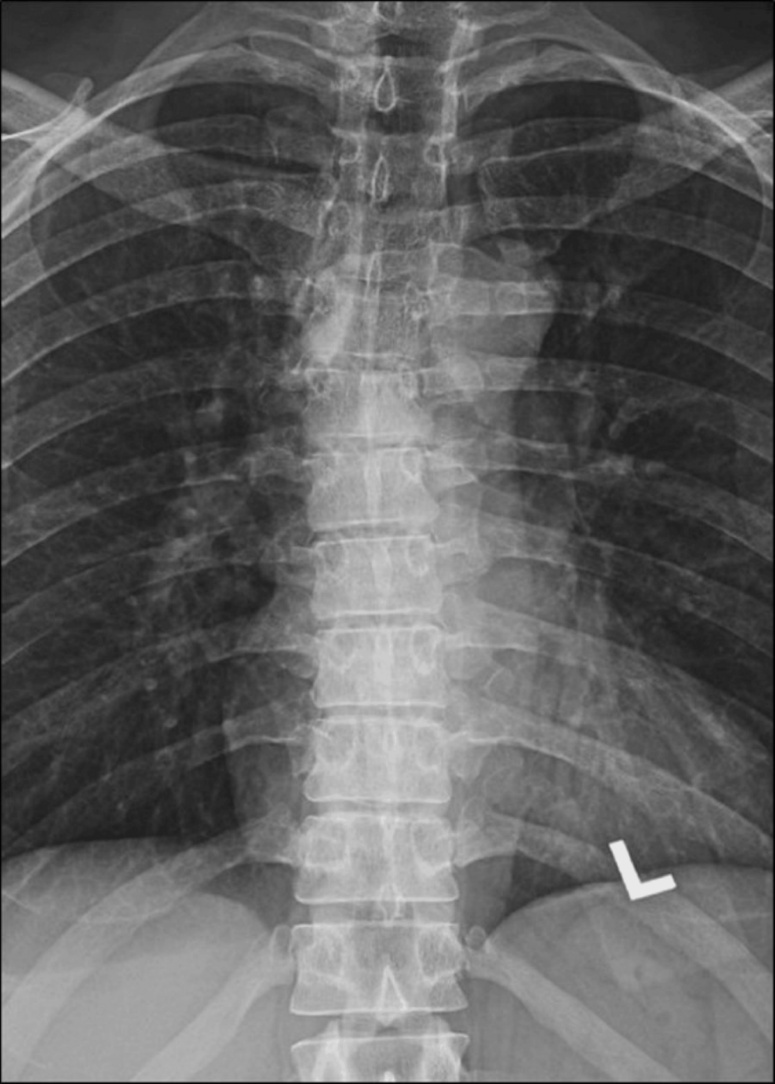
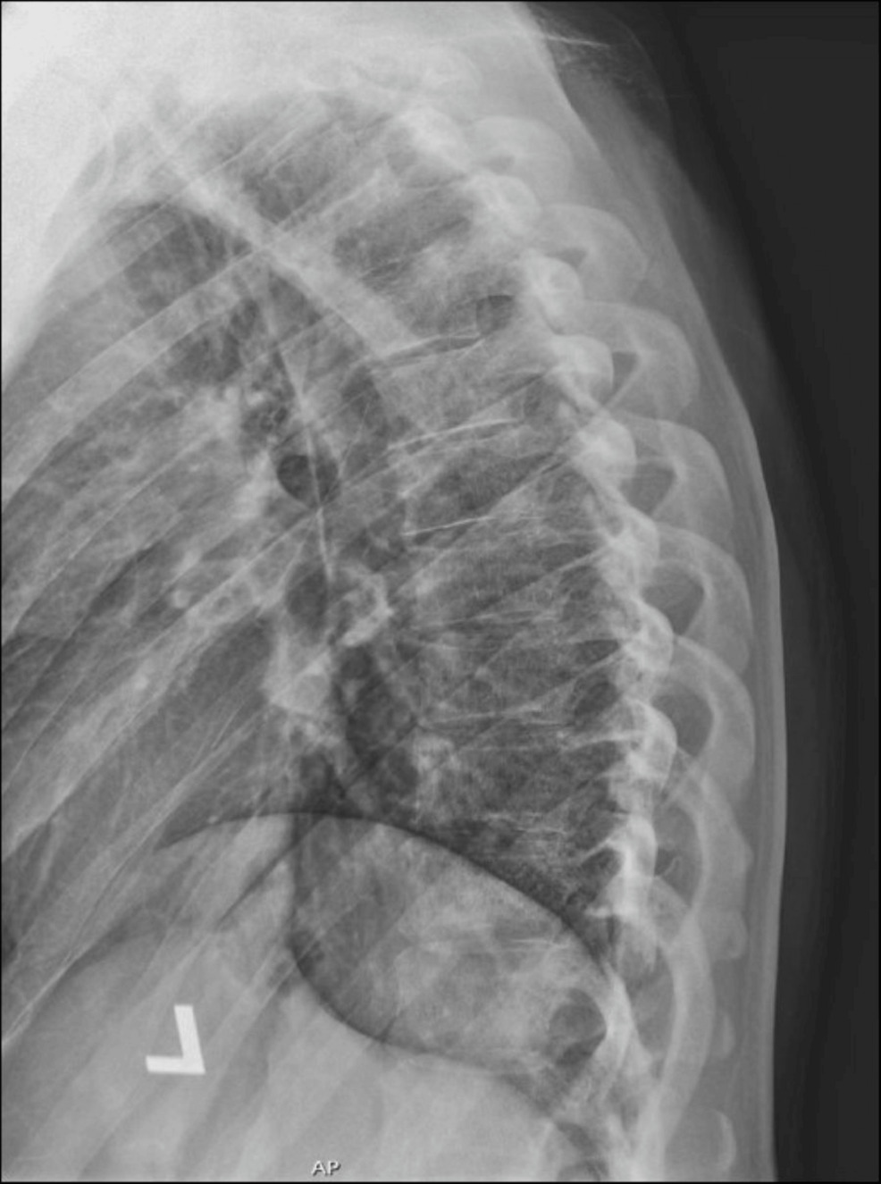
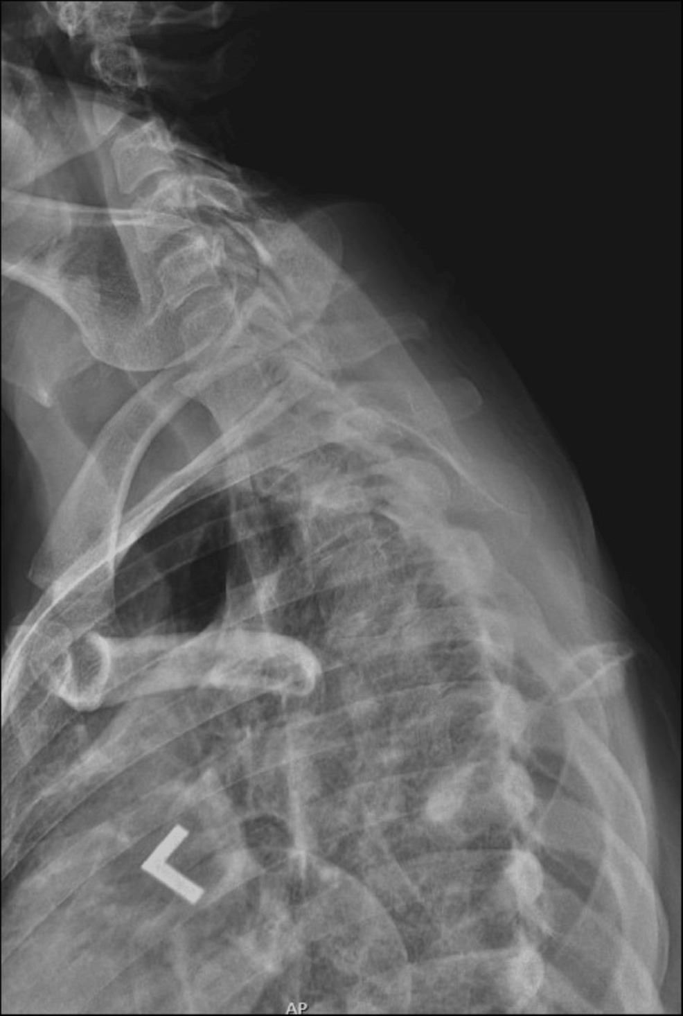
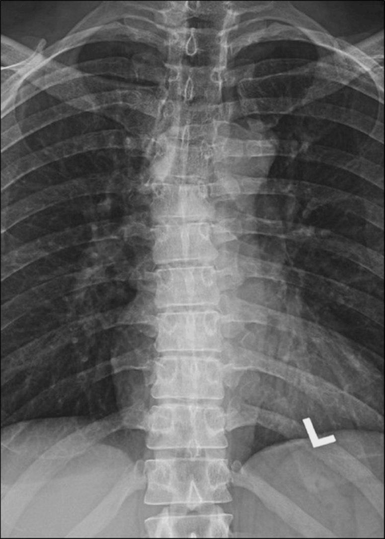
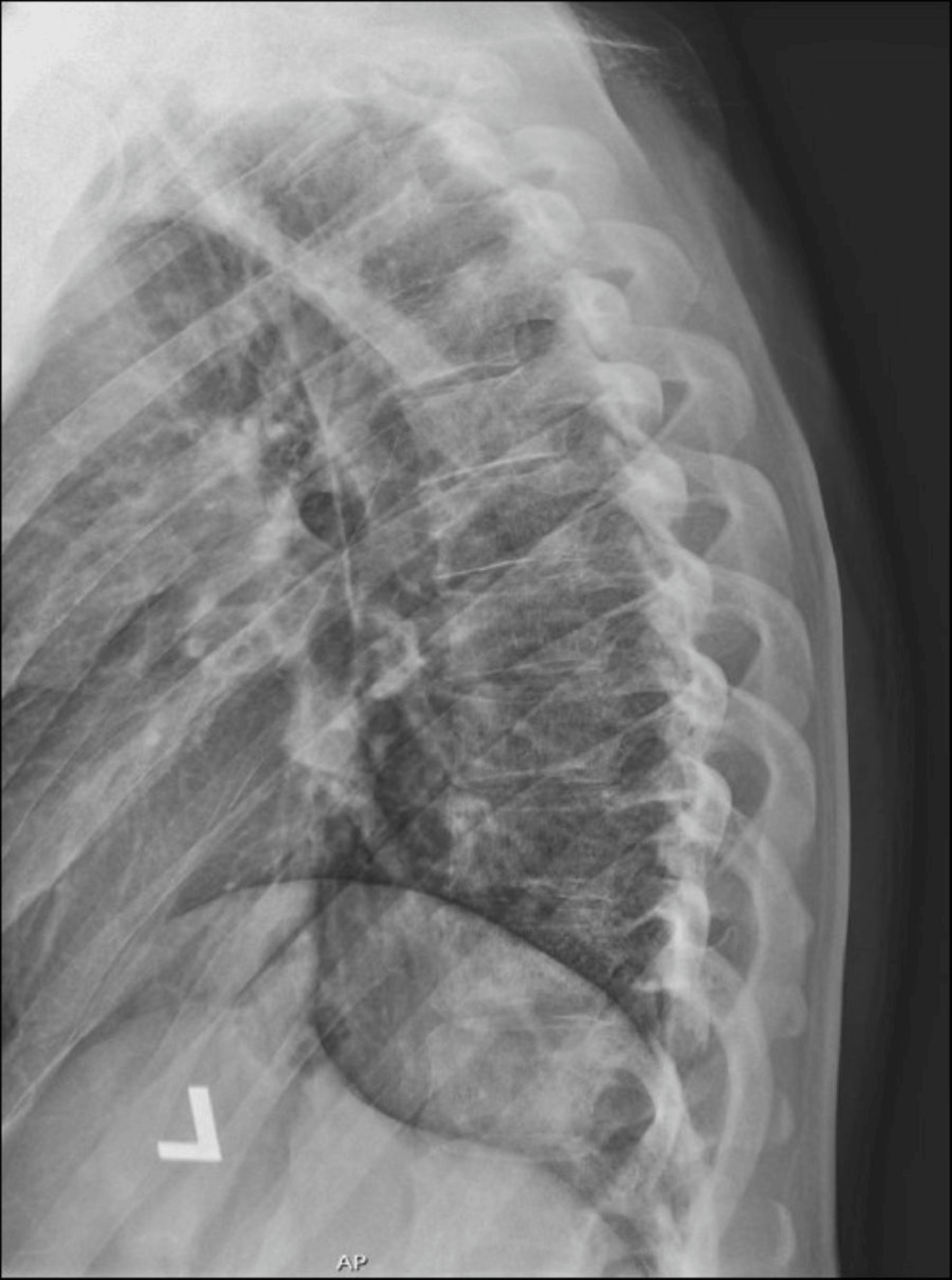
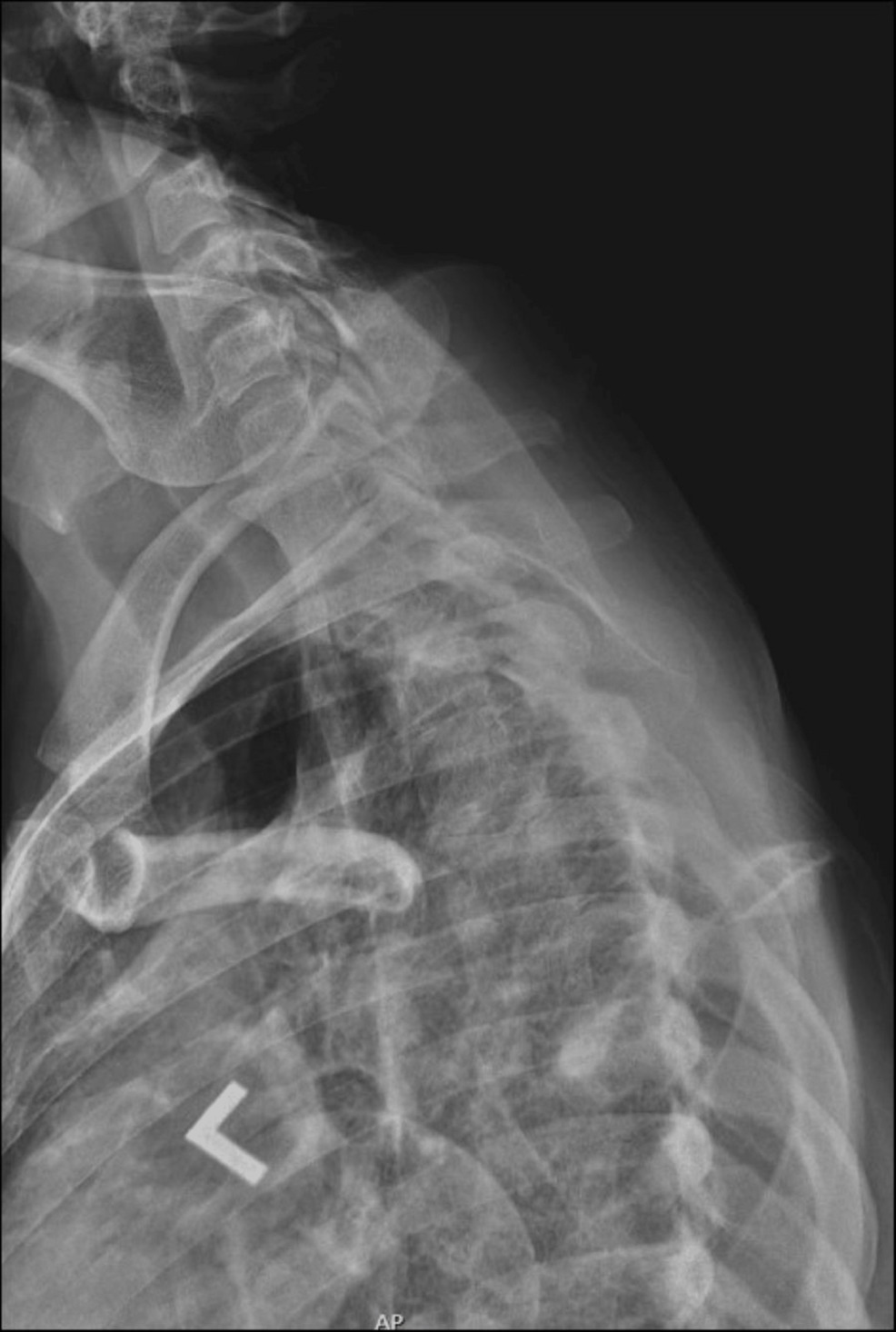
Examination noted a negative Spurling’s test, pain-free cervical flexion/extension, and tenderness over the thoracic spine and surrounding paraspinal muscles. The localized pain was exacerbated by sneezing and deep breathing. There was no lasting pain relief with ice, NSAIDs, heat, or home exercises. He was diagnosed as having thoracic spine pain and was prescribed cyclobenzaprine and corticosteroids. Laboratory testing (inflammatory markers, complete metabolic panel, CBC, PSA level) was ordered but unclear if they were performed, as no lab results were documented in the records. Thoracic radiographs were obtained and interpreted by a medical radiologist as normal. See figures 1-3. Thoracic spine MRI was recommended but not ordered.
The following week, the patient saw a chiropractor for the same chief complaint and was diagnosed with thoracic spine subluxation. The chiropractor performed a supine thoracic spine manipulation, providing the patient with relief for the first time in months.
Only temporary relief was obtained from the manipulation, and the patient sought care with another PCP 2 times in the next month due to ongoing thoracic pain. Cyclobenzaprine and prednisone were prescribed. Laboratory testing (complete metabolic panel, CBC with differential/platelet, ESR, WES, ANA, CRP, complete UA) was performed with no significant results. Thoracic spine MRI was ordered on both visits but not performed.
Within a week of seeing the second PCP, the patient visited a second DC with continued thoracic pain that was not responsive to the prescribed steroids or muscle relaxants. Subjectively, the patient’s pain level had worsened to a consistent 7/10. Despite nightly use of analgesics and heat therapy to aid sleep, his daily functioning had gradually declined since the symptom onset. Upon presentation, the patient denied radiating pain, weakness, or sensory loss in the upper or lower extremities, shortness of breath, or anterior chest wall soreness.
Objectively, active cervical and thoracolumbar spine range-of-motion (ROM) testing revealed diffuse reductions in ROM, with mild pain and stiffness in the upper thoracic region. The Soto-Hall test produced mild, dull aching in the mid-upper thoracic region. The cervical distraction test alleviated most of his mid-upper thoracic pain. Neurologic and muscular examination was unremarkable. Cervical spine radiographs were taken in the chiropractic office and sent to a chiropractic radiologist with DACBR (Diplomate of the American Board of Chiropractic Radiology) credentials for interpretation. Mild discogenic spondylosis and uncovertebral arthrosis at C6/C7 were noted.
The chiropractor’s differential diagnosis was
-
Cervical facet-mediated pain at C6/7 with referral pain into the upper thoracic region.
-
Lower cervical discal pathology.
-
Chronic mid-upper thoracic segmental fixation with associated muscular rigidity secondary to poor posture.
Ten sessions of cervical traction and spinal manipulation of the cervical and thoracic regions were completed with the second DC. In addition, the chiropractor administered ultrasound treatments and performed soft tissue release over the affected regions. Home exercises and stretches provided symptomatic improvement.
On the 10th visit, the patient reported that the original thoracic pain had almost completely resolved; he currently noted intermittent discomfort rated 0-2/10 on the VAS. He was sleeping pain-free and had returned to the gym several times in the past week without issues. On this visit, he also reported mild anterior thigh fatigue or weakness over the past week, which he attributed to his recent lack of exercise. He denied gait issues, difficulty with stairs, or heel/foot drop. Normal gait and heel-toe walk were observed. Active cervical and thoracic spine ROM was full and without pain; orthopedic tests were non-provocative; and neurological testing did not demonstrate any deficits.
On this visit, the differential diagnosis included:
-
Fatigue related to the recent resumption of physical activity after a period of inactivity.
-
Upper motor neuron lesion.
The chiropractor recommended a follow-up visit within 3 to 5 days. The patient was instructed to monitor symptoms and seek medical attention immediately if leg fatigue/weakness progressed.
On the next and final visit, the patient presented with increasing weakness in the lower extremities. He denied any loss of motor control, alterations in bowel or bladder function, lower extremity pain, paresthesia, numbness, or genital hypoesthesia.
The patient reported no thoracic spine pain and had normal thoracolumbar spine ranges of motion. Orthopedic testing revealed the presence of Lhermitte’s Sign. Maximum cervical compression testing produced mild middle-upper thoracic spine pain. Ankle dorsiflexion and big toe extension were graded at 4/5 bilaterally. Beevor’s and Hoffman’s signs were negative, as was Valsalva’s upon testing. Ankle clonus and Babinski’s sign were also negative bilaterally.
The subjective and objective findings were not conclusive but suggested an upper motor neuron lesion secondary to a potential space-occupying lesion. No spinal manipulation was performed, and a referral to a spine surgeon was made, with recommendations for cervical and thoracic MRI imaging.
There was a 6-day delay in surgical consultation due to scheduling issues in the orthopedic surgeon’s office, at which point the second chiropractor contacted the surgeon directly and facilitated a next-day appointment with the surgeon. The patient’s symptoms were persistent at the time of examination, and cervical and thoracic MRI imaging was performed.
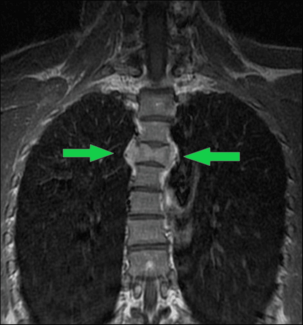
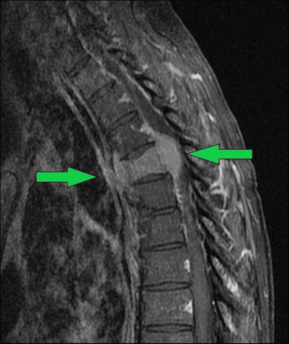
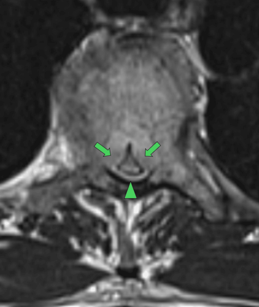
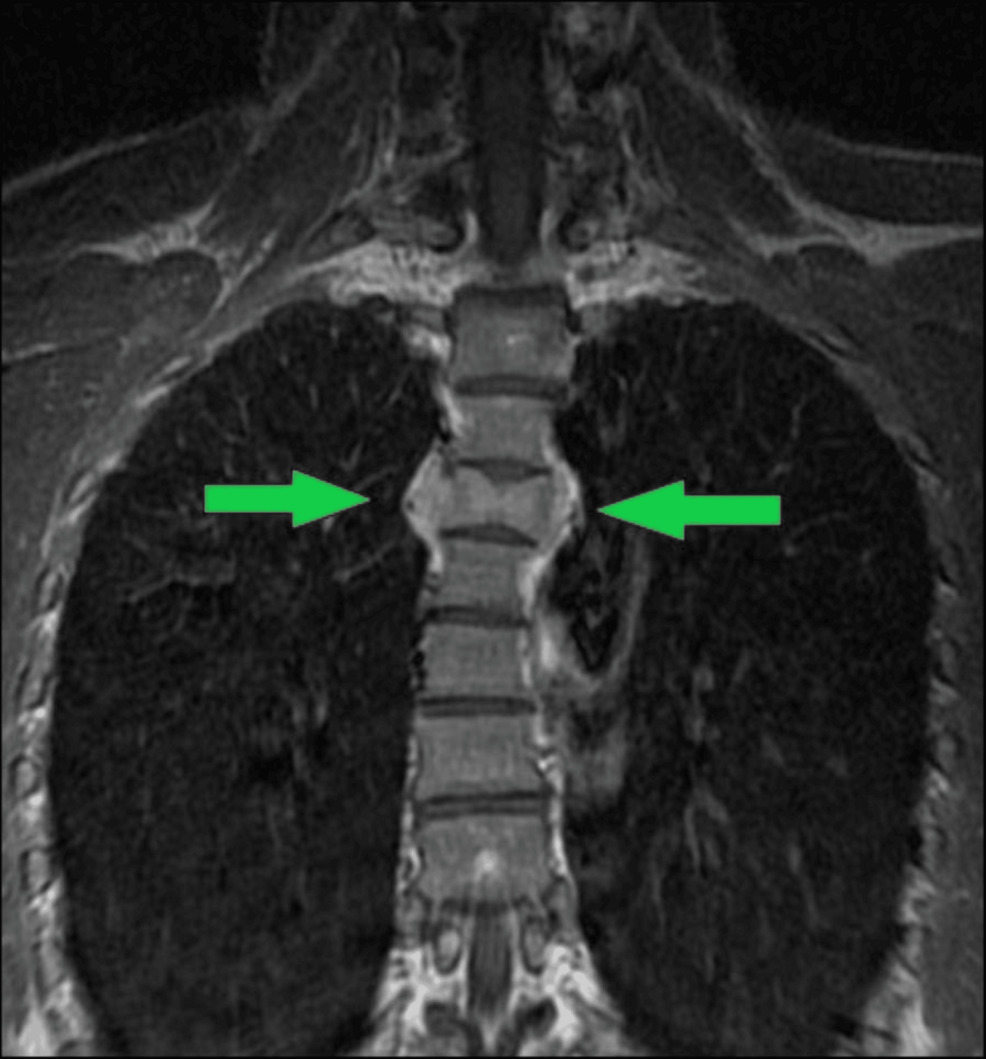
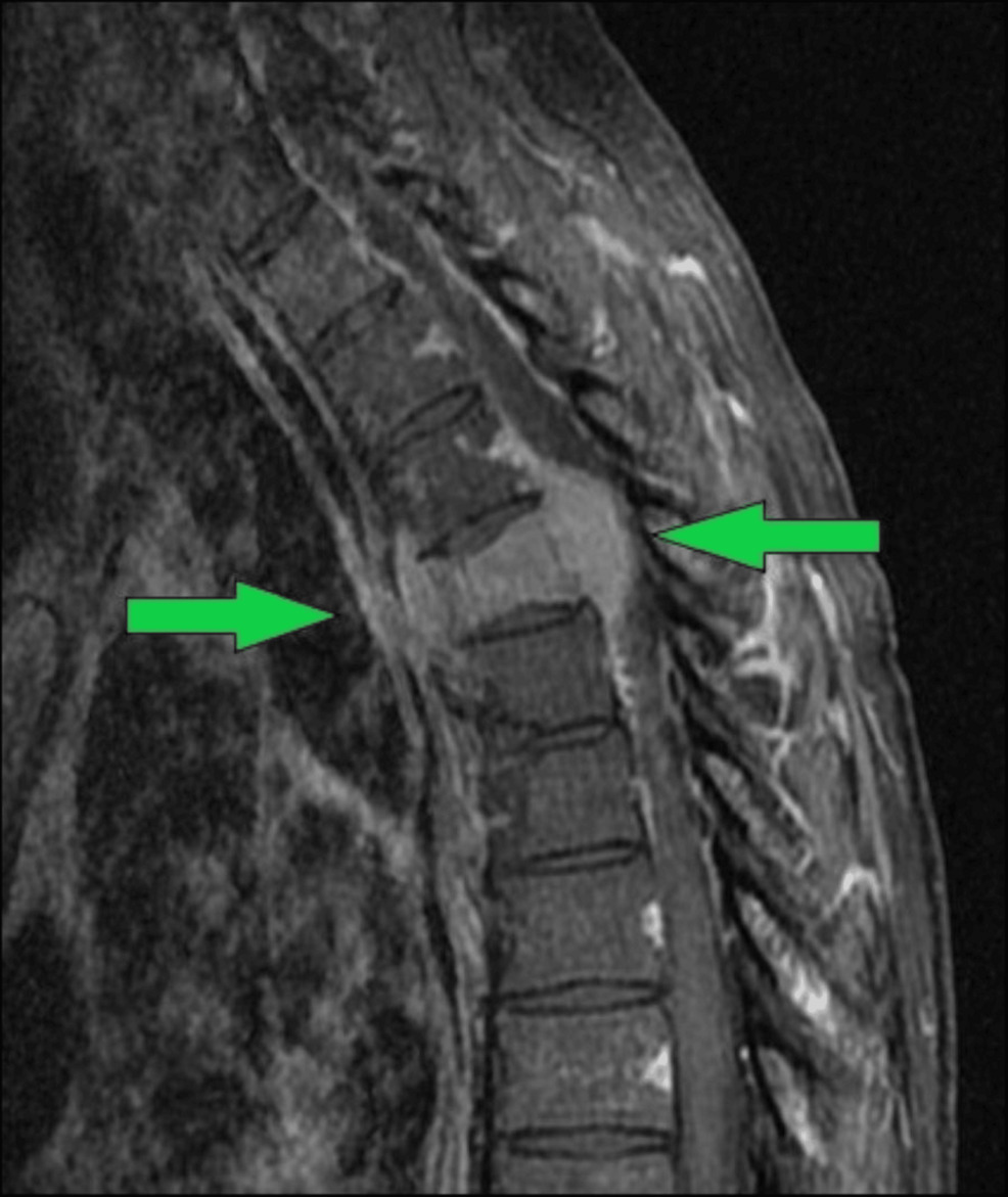
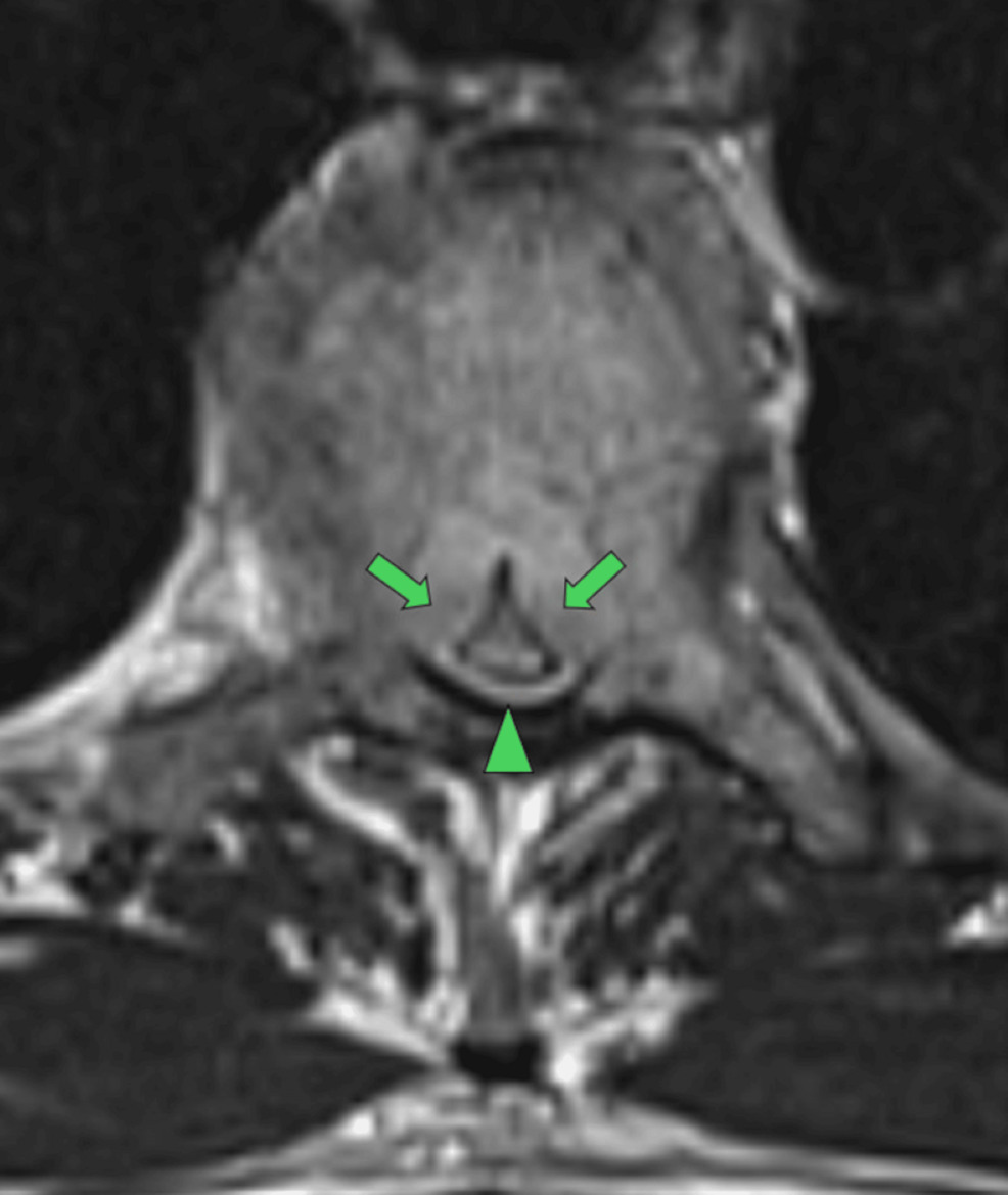
Imaging demonstrated a moderate T4 pathologic compression fracture consistent with a neoplastic process. (Figures 4-6) Retropulsion with extension into the canal caused severe canal stenosis and significant cord compression.
Intervention And Outcomes
Based on the MRI findings, a neurosurgical consult was obtained, and surgery was performed within 2 days. The procedure performed was a partial T4 resection, cage placement, and T2-6 fusion.
Histological testing was performed. The pathology report stated: “The morphological features are those of a neoplastic (clonal) plasma cell proliferation. Neoplastic (clonal) plasma cell proliferations, or plasma cell dyscrasia, can include plasmacytoma, monoclonal gammopathy of uncertain significance (MGUS) or plasma cell (multiple) myeloma.” Clinical and radiological correlation supported the diagnosis of solitary bone plasmacytoma of the T4 vertebral segment. Photos of the pathological analysis of the specimen were not available for review.
Radiation therapy was administered for 4-5 months, and chemotherapy several times per week. At the time of this report, the patient reported current remission following a successful stem cell transplant in the Fall of 2025, and was able to return to work and most activities of daily living. Case events are summarized in Table 1.
DISCUSSION
This case report describes a 42-year-old male who presented with progressive thoracic spine pain that failed to resolve with primary care management and chiropractic intervention. Despite near-resolution of thoracic spine pain following targeted cervical and thoracic treatment from a second chiropractic intervention, the subsequent emergence of new neurological symptoms prompted timely reassessment, referral, and MRI imaging that led to a diagnosis of T4 solitary bone plasmacytoma.
Other causes of thoracic pain that may mimic benign neuromusculoskeletal causes include, but are not limited to, acute myocardial infarction, neoplasms, infections, aortic dissections, and viscerally referred pain.12 Additional differential diagnoses for thoracic pain include thoracic disc lesions, fractures, and Scheuermann’s disease.12
This case highlights the importance of ongoing clinical vigilance, serial history-taking, and red-flag screening in clinical practice, as well as the value of prompt referral and interprofessional collaboration to prevent diagnostic delay in rare but serious pathology.13–18 Thoracic spine pain is a common somatic complaint that presents to chiropractic physicians and other allied health providers, making clinical attentiveness vital on every patient interaction.
Differential diagnosis in this case before MRI imaging was challenging due to the rare nature of this plasmacytoma. Even though the 2nd chiropractic intervention relieved the patient’s persistent thoracic spine pain for the first time in several months, somatic pain related to these conditions can fluctuate, making any improvement short-lived. The clinical improvement supported the initial differential diagnoses rather than suggesting alternative causes due to a lack of clinical progress. Practitioners should remain attentive to other changes in the clinical presentation, regardless of symptom relief.
Without the subsequent neurological changes, this diagnosis would have been more challenging. While such conditions are rare, they underscore the importance of considering the entire clinical picture and monitoring patients for clinical changes. Even near-complete resolution of nociceptive symptoms with conservative care does not rule out serious underlying pathology.
Another key lesson highlighted by this case is the importance of ensuring completion of recommended or ordered laboratory testing and advanced imaging. The 1st PCP ordered lab testing and recommended a thoracic spine MRI, while the 2nd PCP subsequently ordered the thoracic spine MRI twice. However, the first lab order and the thoracic spine MRI examination were not performed. Review of the medical records showed no evidence of follow-up by either PCP office regarding the status of those orders and recommendations.
Retrospective, blinded reviews of the December 2024 thoracic radiographs by two chiropractic radiologists and a medical radiologist found no abnormalities and confirmed the initial radiology report.
Limitations
This case report is limited by its single-patient design, which restricts generalizability to other patients with similar presentations. It also lacks long-term follow-up data. As an observational report without a control group, it cannot determine the specific effect of chiropractic care on the patient’s significant decrease in pain or overall outcome.
CONCLUSION
This case highlights the limitations of orthopedic testing in diagnosing specific conditions and underscores the importance of exploring the causes of other symptoms (progressive leg weakness) which occur even when near-complete symptomatic pain relief is obtained. In this instance, diligent oversight of symptom variations and subsequent examinations demonstrated the progression of the clinical condition over time. MRI imaging ultimately identified the condition after plain radiographs failed to detect the lesion three months earlier.
Notably, the patient’s pain was nearly resolved after the second chiropractic intervention, but the underlying pathology eventually progressed, altering the clinical picture. Temporary improvement of nefarious conditions has been documented in the natural history of solitary bone plasmacytoma, prior to definitive diagnosis.
We suggest that the solitary bone plasmacytoma at T4 produced secondary musculoskeletal pain through its effects on nearby soft tissues, paraspinal muscles, and surrounding structures. This secondary neuromusculoskeletal pain responded favorably to conservative chiropractic treatment. However, the neoplastic lesion continued to advance independently, eventually weakening the vertebral body.
The delayed onset and subsequent worsening of neurological symptoms were most likely caused by pathologic collapse of the T4 vertebra, resulting in retropulsion of bone fragments, spinal canal narrowing, and compression of the spinal cord. It emphasizes the need for heightened clinical vigilance, timely re-evaluation when symptoms change or fail to resolve fully, and consideration of advanced imaging when red-flag features develop.
Informed consent
Written informed consent was obtained from the patient for the publication of this case report.